SDT fatty rat, spontaneously type 2 diabetes animal model, shows the symptoms of secondary sarcopenia related with diabetes and obesity.
According to the pathophysiological findings of the skeletal muscle related with glycolipid metabolism disorder indicated of secondary sarcopenia, SDT fatty rat should be considered to be a useful model for analysis of diabetes-related sarcopenia.
▼ Inquiry here
Method
(Click on the image to see a larger image)
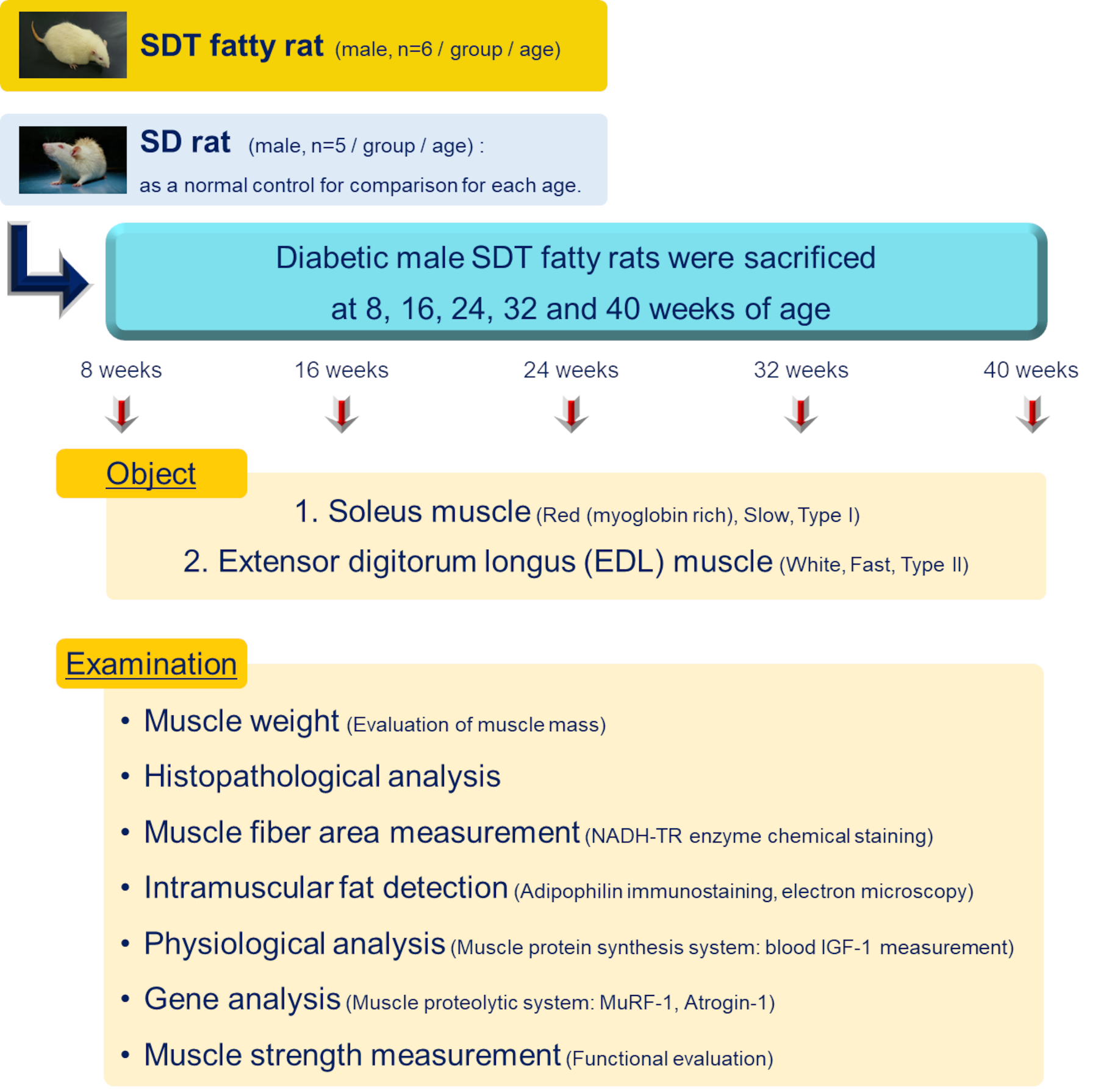
Animals
・Spontaneously Diabetic Torii Lepr fatty (SDT fatty) rat (male, n=6/group/age)
・Sprague-Dawley (SD) rat (male, n=5/group/age): as a normal control for comparison for each age.
Anatomy / Coverage
Diabetic male SDT fatty rats were sacrificed at 8, 16, 24, 32 and 40 weeks of age
Object
1. Soleus muscle (Red (myoglobin rich), Slow, Type I)
2. Extensor Digitorum Longus (EDL) muscle (White, Fast, Type II)
Examination
・Histopathological analysis
・Muscle fiber area measurement (NADH-TR enzyme chemical staining)
・Intramuscular fat detection (Adipophilin immunostaining, electron microscopy)
・Physiological analysis (Muscle protein synthesis system: blood IGF-1 measurement)
・Gene analysis (Muscle proteolytic system: MuRF-1, Atrogin-1)
・Muscle strength measurement (Functional evaluation)
Statistical analysis Statistical analysis
Student t-test or Aspin-Welch t-test
Result
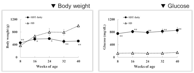
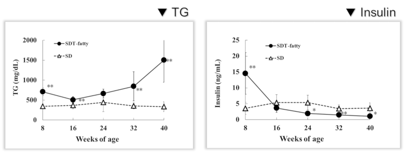
Result 1: Body weights and biochemical parameters
SDT fatty rat mean body weight was 23% higher than that of SD rats at 8 weeks of age but was lower than that of SD rats from 16 weeks of age thereafter. Serum glucose, TG, and TC levels were significantly higher than those in the SD rats at 8 weeks of age. Serum insulin levels were markedly higher at 8 weeks of age and were sharply decreased from 16 weeks of age.
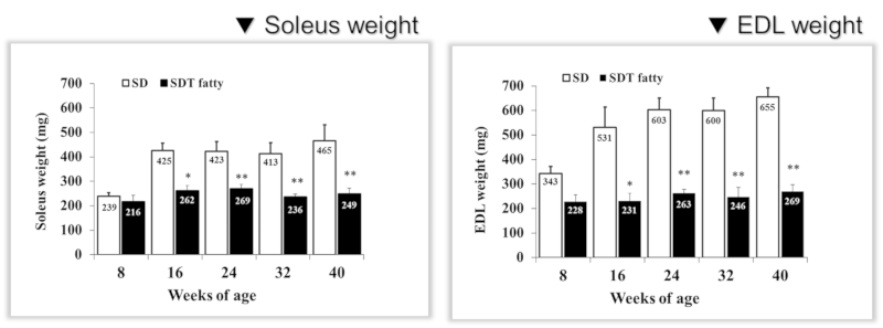
Result 2: Skeletal muscle weight (muscle mass)
Muscle weights of the soleus and EDL were significantly lower in the SDT fatty rats than those in the SD rats from 16 weeks of age. The EDL tended to be decreased more than soleus.
Muscle weights (absolute/relative (per BW)) of SDT fatty rats at 16 weeks of age were 39.4%/29.7% lower in the soleus and 57.5%/50.6% lower in the EDL than those of SD rats.
SD rats showed age-dependent increases in the weights of both muscles 1.9 times higher between 8 and 40 weeks of age. On the other hand, the weights of both muscles in SDT fatty rats were almost constant to 40 weeks of age. This showed the aging-dependent increases of the skeletal muscle of SDT fatty rat was significantly lower than the normal control.
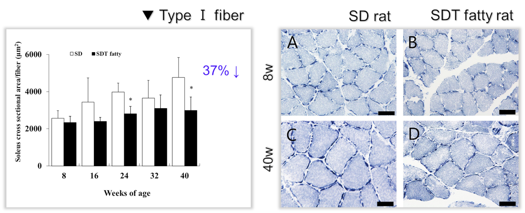
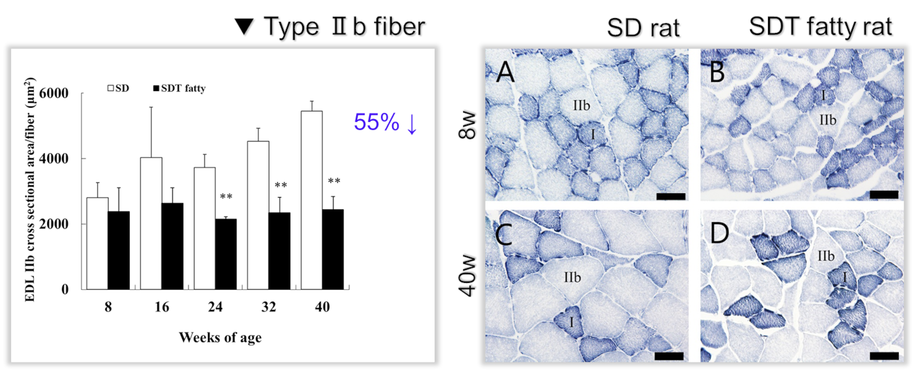
Result 3: Pathological analysis and cross-sectional area measurement of skeletal muscle
There was no pathological significant difference between both SDT fatty and SD rats in 8 weeks of ages. Then SDT fatty rat showed 30% decrease of cross-sectional area in both soleus and skeletal muscles from 16 weeks of age. From 24 weeks of ages, SDT fatty rat showed statistical decreasing in cross-sectional area of both muscles.
The mean cross-sectional areas of the soleus muscle fibers (mainly composed of type I fiber) of the SDT fatty rats were 8.9%, 30.0%, 29.4%, 15.2% and 37.2% lower than those of the SD rats at 8, 16, 24, 32 and 40 weeks of age, respectively.
On the contrary, the mean cross-sectional areas of the type IIb fibers in the EDL of the SDT fatty rats were 15.4%, 34.6%, 42.3%, 48.2% and 55.3% lower than those of SD rats respectively. However, type I fibers in the soleus were not meaningful difference.
SDT fatty rat showed less age-related development of the cross-sectional area of the skeletal muscle than SD rat during 8 to 40 weeks of age. This decrement was shown the type II muscle fiber more than the type I.
(Click on the image to see a larger image)
(Click on the image to see a larger image)
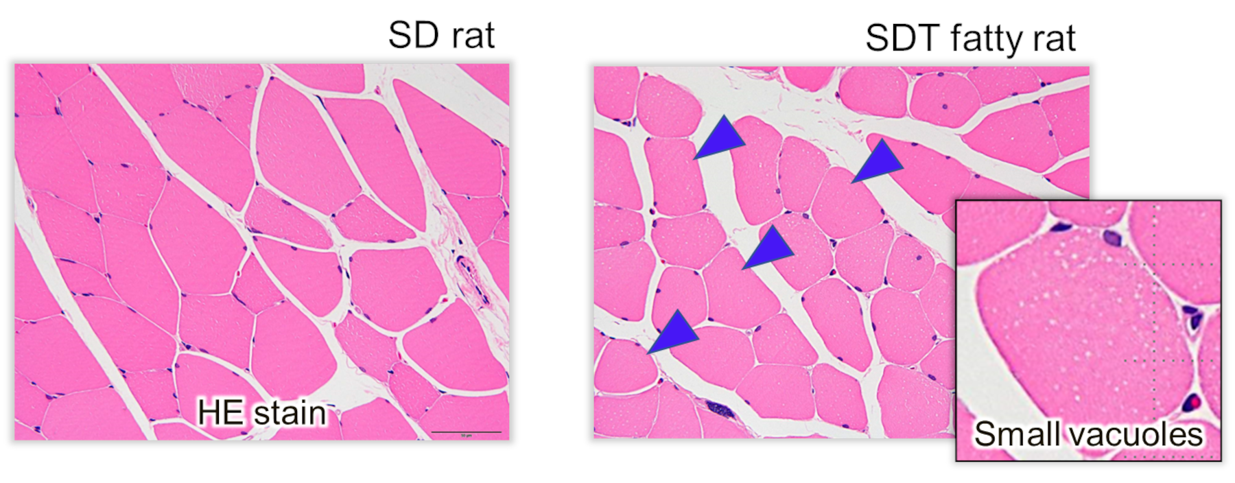
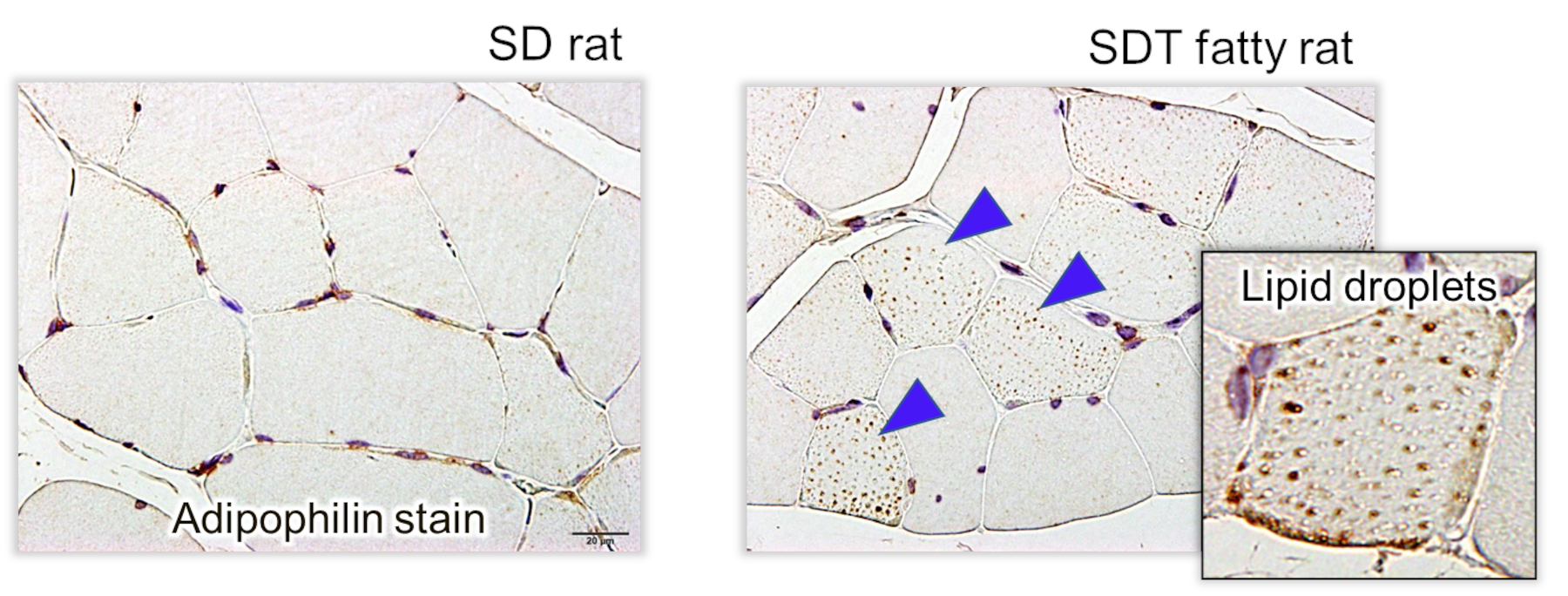
Result 4: Intramyocellular lipid (IMCL) droplet accumulation and disrupted mitochondria
By HE stains analysis, small vacuoles in the muscle fibers were increased in the soleus and EDL in the SDT fatty rats from 8 weeks of age. Intramuscular vacuoles were also observed in the SD rats but there were fewer numbers compared with in the SDT fatty rats at the same age. The vacuoles showed positive on adipophilin immunohistochemistry, a marker of lipid protein.
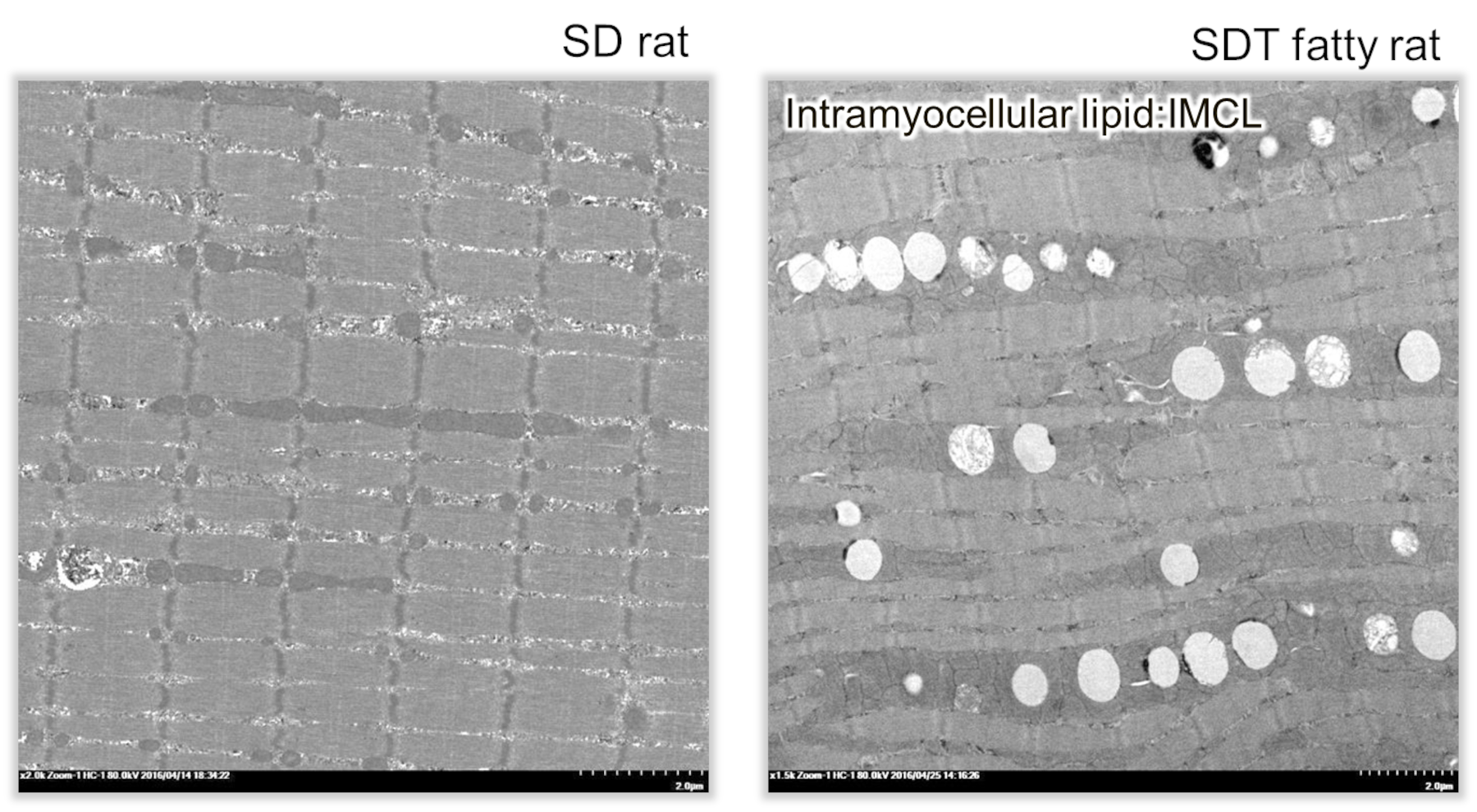
So that these vacuoles were identified as lipid droplets. In addition, lipid droplets were characterized by low electron dense bodies in longitudinal sections seen on electron microscopy.
Lipid droplets of the skeletal muscle were located its intermyofibrillar mainly and close to its mitochondria. There were some mitochondria showing disrupted internal structures.
(Click on the image to see a larger image)
(Click on the image to see a larger image)
(Click on the image to see a larger image)
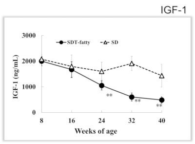
Result 5: Serum IGF-1 level
Plasma IGF-1 level of the SDT fatty rat was comparable to that of the SD rat for up to 16 weeks of age, however, sharply decreased from 24 weeks of age.
About plasma IGF-1, the SD rat showed the level continuously from 8 weeks of age, SDT fatty rat showed decreasing 50% to 24 weeks of age, and 75% to 40 weeks of age.
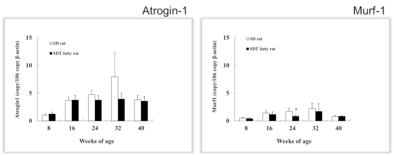
Result 6: Gene expression analysis of the muscle protein resolution
Messenger RNA levels of Atrogin-1 and Murf-1 in the EDL, which was changed more than soleus in muscle weight and pathological analysis, were measured as the parameter of the muscle protein resolution.
It was expected that both gene expression increased if resolution was accentuated. However, both of all aged examined SDT fatty were not increased.
In this study, these proteolysis markers did not increase in SDT fatty rats at any of the ages examined.
Result7: Forelimb grip strength test for muscle strength
For measuring skeletal muscle function, examination of muscle strength was conducted using a Grip Strength Meter (GPM-100B, Malmquist, Toyama, Japan) to evaluate muscle function of SDT fatty rats in 8, 24, and 40 weeks of age. There were no differences in mean score for the grip strength test between SD rats and SDT fatty rats at 8 weeks of age.
But significant decreases in muscle strength were observed in the SDT fatty rats at 24 and 40 weeks of age. The mean scores for the muscle strength test in the SDT fatty rats were 32% and 38% lower at 24 and 40 weeks of age, respectively, compared with those of the SD rats.
SDT fatty rat showed skeletal muscle function decreasing in 24 and 40 weeks of age.
Manuscript:
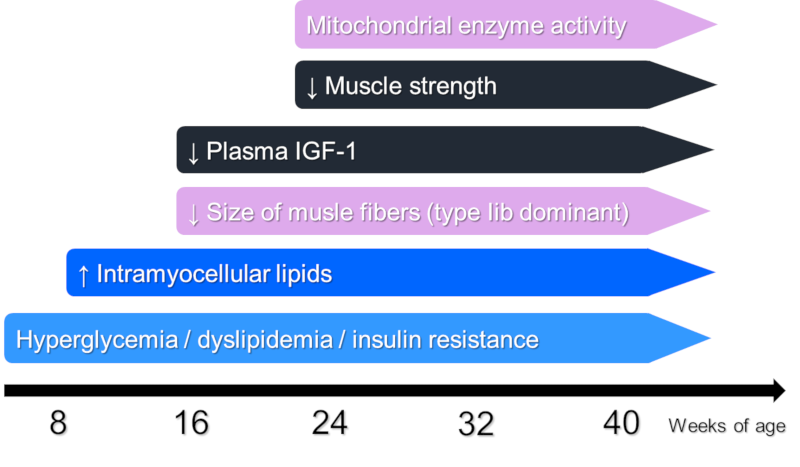
SDT fatty rat showed sarcopenia indication changings such as muscle decreasing, fiber amyotrophia, function decreasing, and protein synthesis signal decreasing comparably more than SD rat. Especially the type 2 fiber-superior amyotrophy was consistent the report of that of human aged sarcopenia.
The accumulation of the lipid droplets inside the skeletal muscle cell was recognized to be related with incline resistance, decreasing protein synthesis, obesity, and diabetes. It could be considered the accumulation of lipid droplets in SDT fatty rat is affected to skeletal muscle atrophy.
▼ SDT fatty rats product page
Inquiry
If you have any question, please feel free to contact us from below.