Retinal lesions were observed in 24-week-old SDT fatty rats, including the retinal thickness, dilatation of the retinal capillaries, decreased pericyte/endothelial cell ratio, and expression of leukocyte adhesion molecules in the retinal capillaries.
SDT fatty rats are highly useful animal model for research on the pathology of human diabetic retinopathy.
SDT fatty rats, which are a type of non-obese type 2 diabetes mellitus (T2DM) model animal that were created by introducing a mutation of the leptin receptor, a gene for obesity, into SDT rats, develop T2DM earlier than SDT rats. As a result, they exhibit diabetic complications such as nephropathy and neuropathy early on. However, there have been only a few reports on diabetic eye complications in SDT fatty rats.
To date, the following results have been revealed in SDT fatty rats.
▼ Inquiry here
Inflammatory-related molecules are increased in the retina of SDT fatty rats.
Real-time PCR
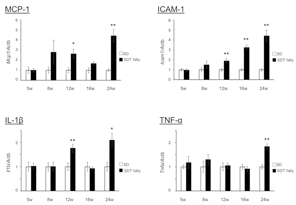
(SD n=5, SDT fatty n=4 , *P<0.05 **P<0.01)
(25th Annual Meeting of The Japanese Society of Ophthalmic Diabetology, 2019)
Luminex assay
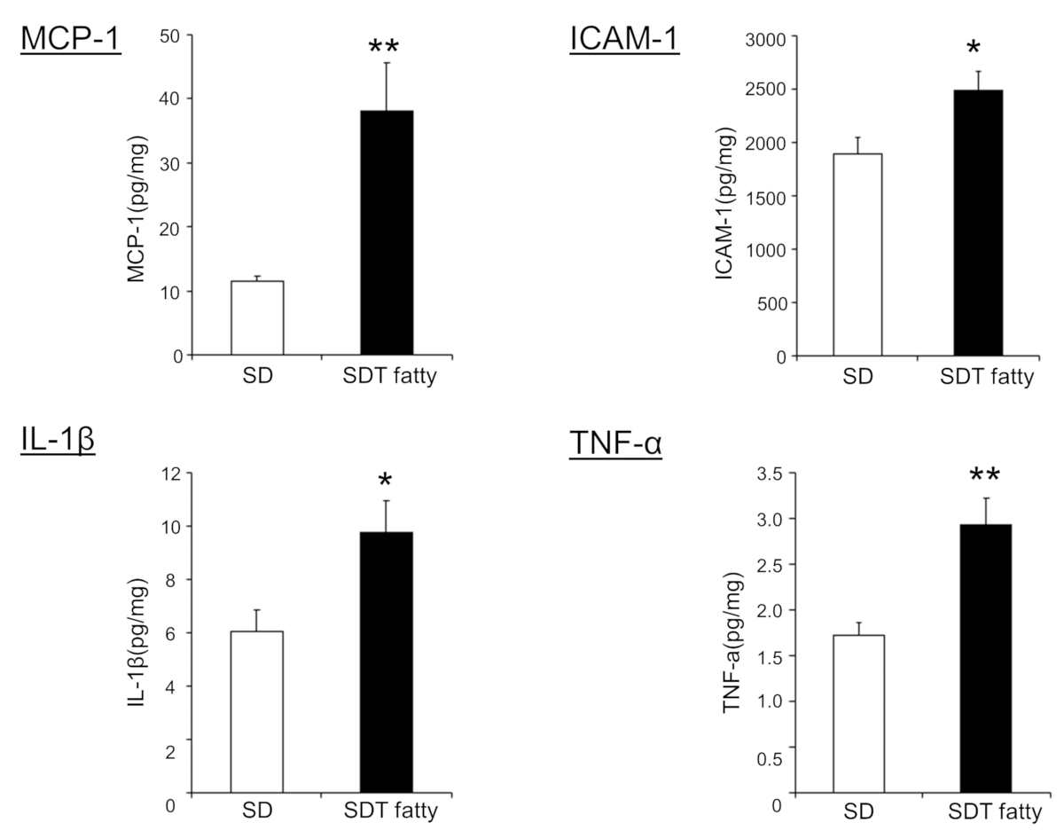
(SD n=5, SDT fatty n=4 , *P<0.05 **P<0.01)
(25th Annual Meeting of The Japanese Society of Ophthalmic Diabetology, 2019)
The purpose of this study was to observe the morphological changes of diabetic eye complications in SDT fatty rats and to contribute to the understanding of the pathogenesis.
Method
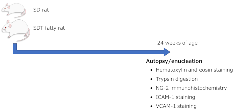
This study investigated the morphological changes of diabetic eye complications in SDT fatty rats. Sprague Dawley (SD) rats were used as the control group.
For the morphological changes of the retina, retinal thickness was measured. The changes in the retinal capillaries were investigated using the following two methods:
- Blood vessels were prepared using the trypsin digestion method, and the capillary diameter and pericyte/endothelial cell ratio were measured.
- The expression of leukocyte adhesion molecules in the retinal capillaries was confirmed by immunohistochemistry.
The results showed that SDT fatty rats had increased retinal thickness, dilated capillary diameter, decreased pericyte/endothelial cell ratio, and increased expression of leukocyte adhesion molecules.
Result
HE staining image (24 weeks old)

Retinal thickness (24 weeks old)
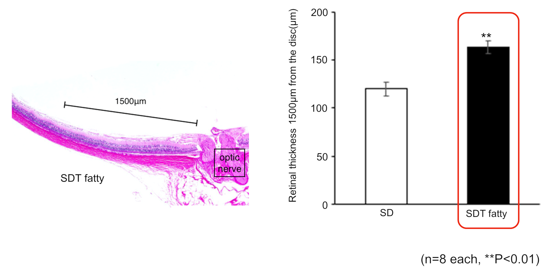
The retina of SDT fatty rats was significantly thicker than that of SD rats.
Triton digestion method
- Fix the extracted eyeballs in 10% neutral formaldehyde.
- Separate the retina and wash it with distilled water overnight.
- Treat with 1% Triton X−100 for 20 minutes.
- Incubate in 3% trypsin solution at 37°C for 60 minutes.
- Separate the inner limiting membrane and non-vascular retinal tissue from the retina in distilled water and isolate the retinal vessels.
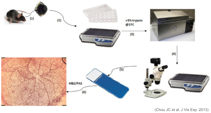
Trypsin-digested specimen (HE staining/24 weeks old)
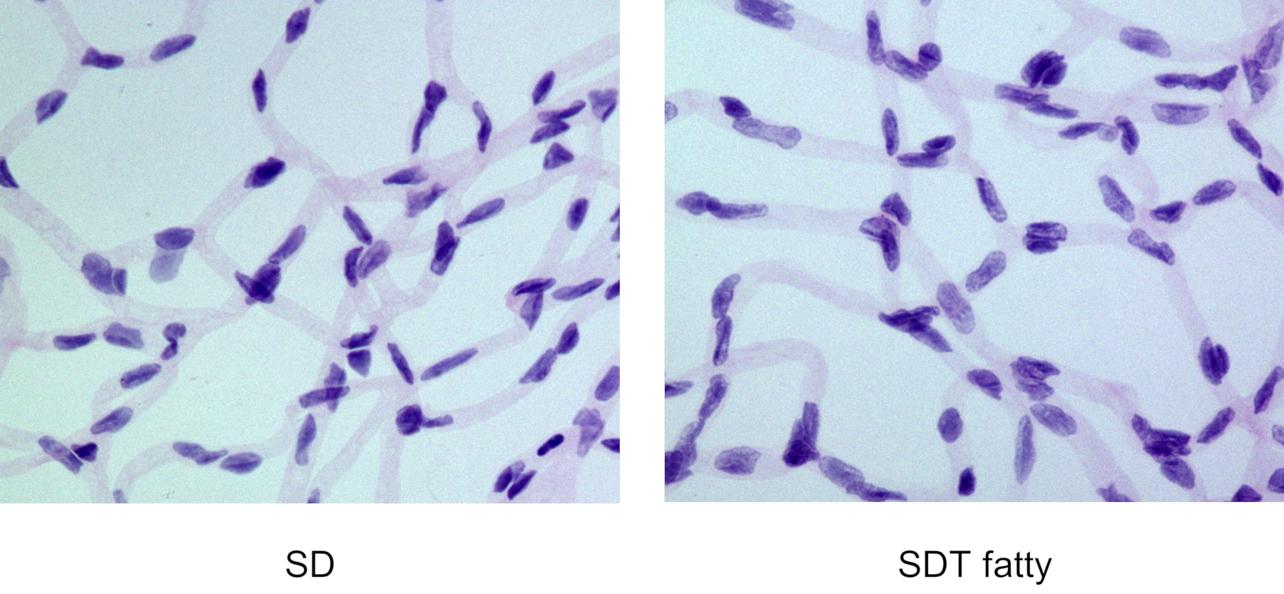
Retinal capillary diameter (24 weeks old)
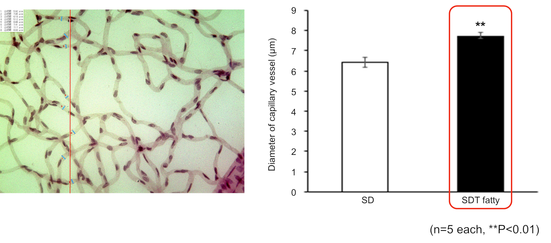
The diameter of the retinal capillaries in SDT fatty rats was significantly dilated.
Pericyte/endothelial cell ratio (24 weeks old)
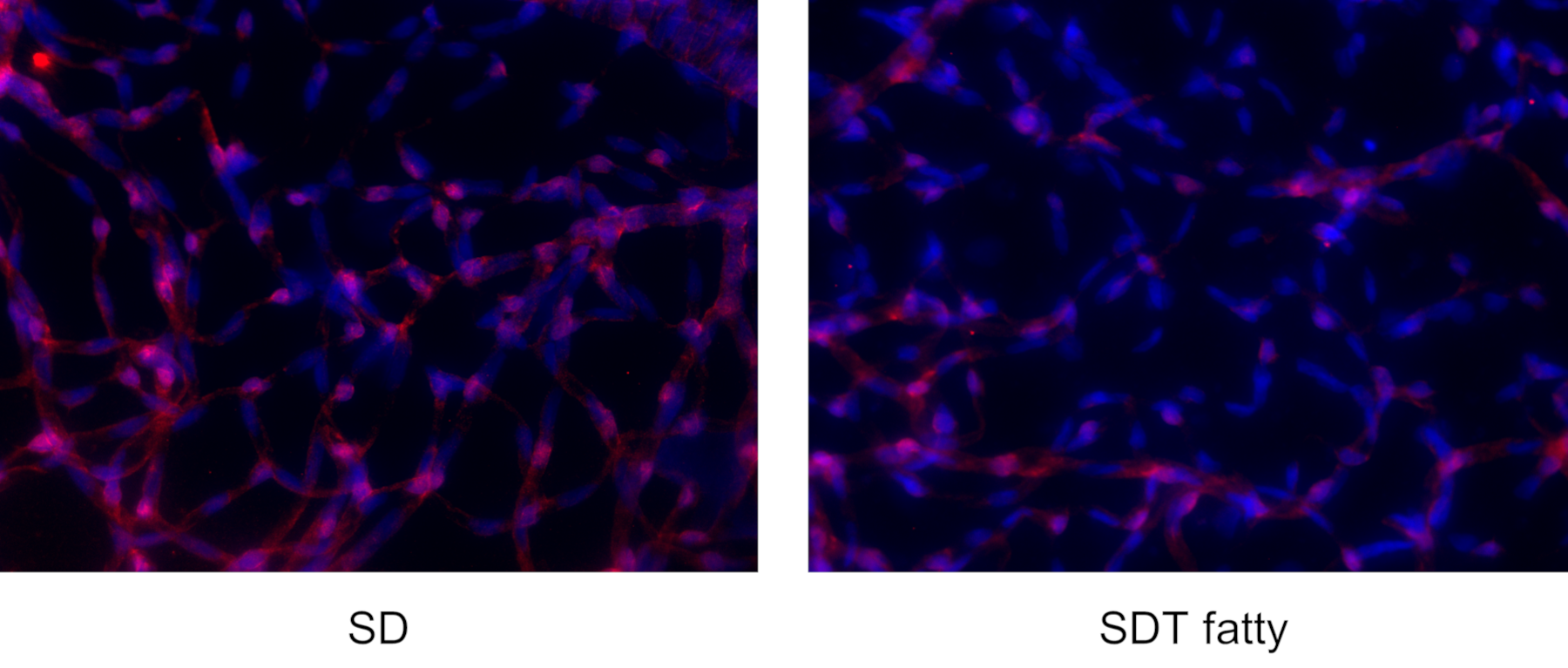
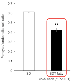
The diameter of retinal capillaries in SDT fatty rats was significantly decreased.
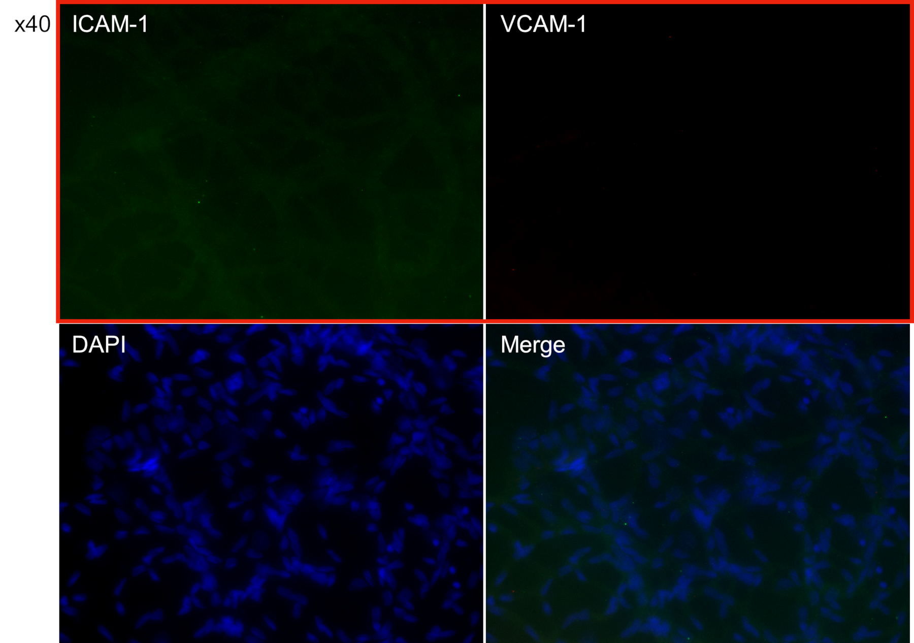
Expression of leukocyte adhesion molecules
Immunostaining (SD rats)
Immunostaining (SDT Fatty rats)
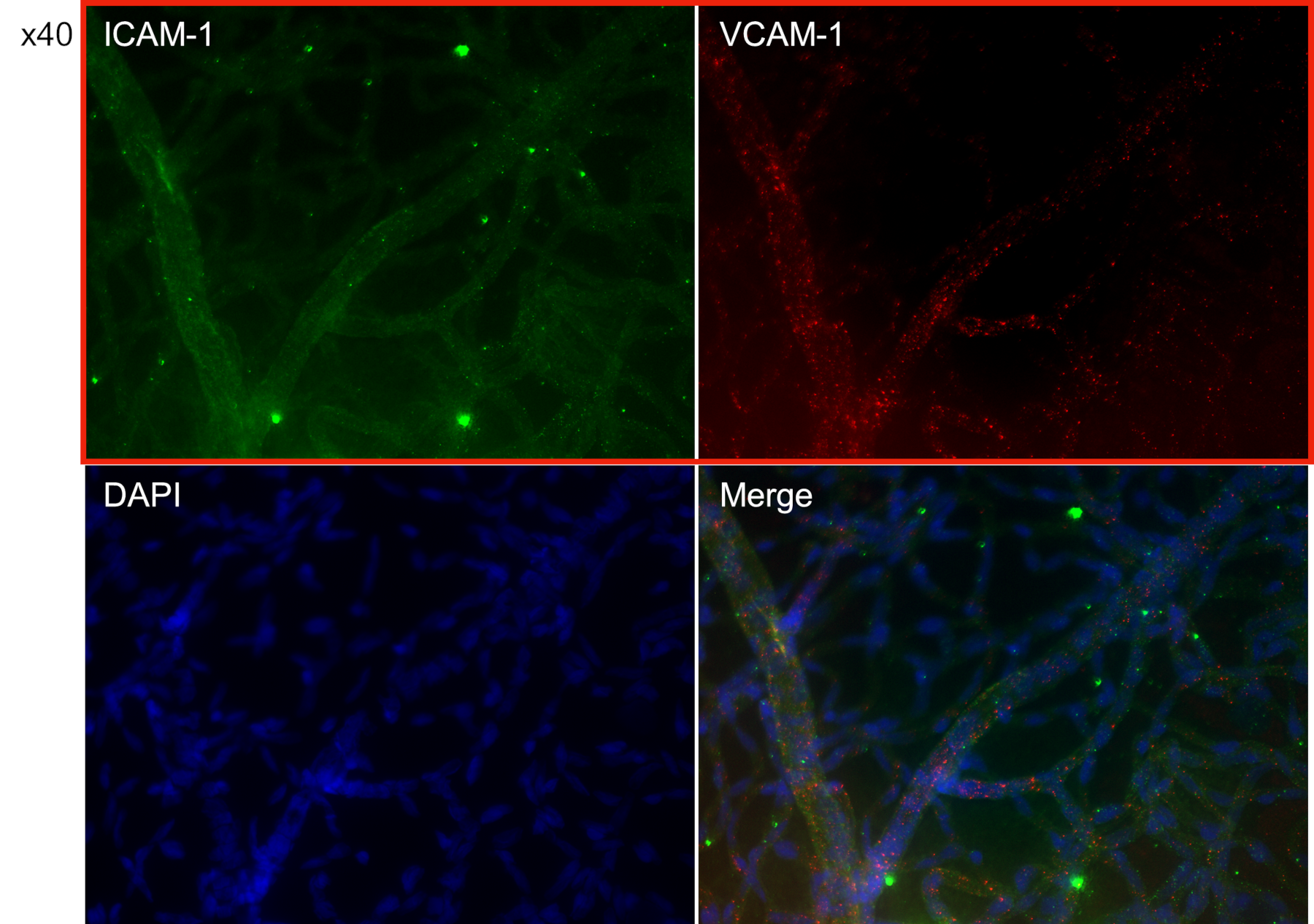
- The expression of leukocyte adhesion molecules ICAM-1 and VCAM-1 was observed in the retinal capillaries of SDT fatty rats.
These results suggest that the following changes occur in the retina and retinal capillaries of SDT fatty rats as diabetes progresses.
Conclusion/Discussion
SDT fatty rats are thought to reflect the early changes of human diabetic retinopathy and are therefore considered to be a useful animal model for the study of the pathogenesis of human diabetic retinopathy.
Early changes of capillaries in diabetic retinopathy
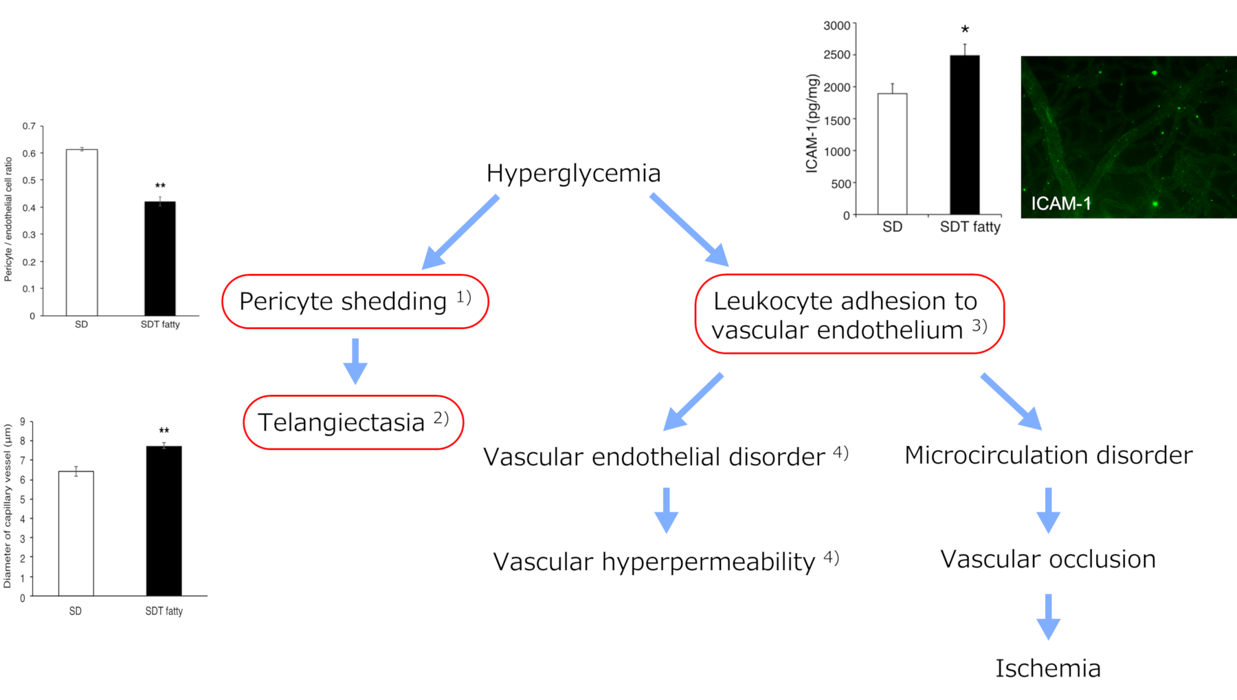
SDT fatty rats reflect the early changes of diabetic retinopathy.
1)Romeo G et al. Diabetes. 2002
2)Dong A et al. Transl Vis Sci Technol. 2021
3)Miyamoto K et al. Proc Natl Acad Sci USA. 1999
4)McLeod D S et al. Am J Pathol. 1995
Specifically:
- As diabetes progresses, various morphological changes occur in the retina and retinal capillaries.
- These changes are like the early changes of human diabetic retinopathy.
- SDT fatty rats can replicate the pathophysiological features of human diabetic retinopathy.
FAQ
When does retinal damage appear?
Product page:
Inquiry
If you have any question, please feel free to contact us from below.