Peripheral neuropathy due to diabetes and drug effects of SGLT inhibitor (Phlorizin) can be observed. Introduction of CLEA Japan's type 2 diabetes model, SDT fatty rats
Spontaneously Diabetic Torii (SDT) Fatty rat is useful animal model to investigate diabetic complications, because SDT fatty rat shows three major microvascular complications of diabetes in kidneys, eyes, and nerves from relatively younger age. Various animal models of diabetes peripheral neuropathy (DPN) have been reported, while there are few reports using SDT fatty rats for DPN.in addition, there have been few reports that perform histopathological analysis of type 2 diabetes animal models.
In the present study, we characterized the DPN in SDT fatty rats by functional and morphological analysis. Blood glucose levels were strictly controlled by treatment of phlorizin (PZN), a non-selective sodium-glucose co-transporter (SGLT) inhibitor to evaluate the effect of hyperglycemia on nerve dysfunction.
▼ Inquiry here
Method
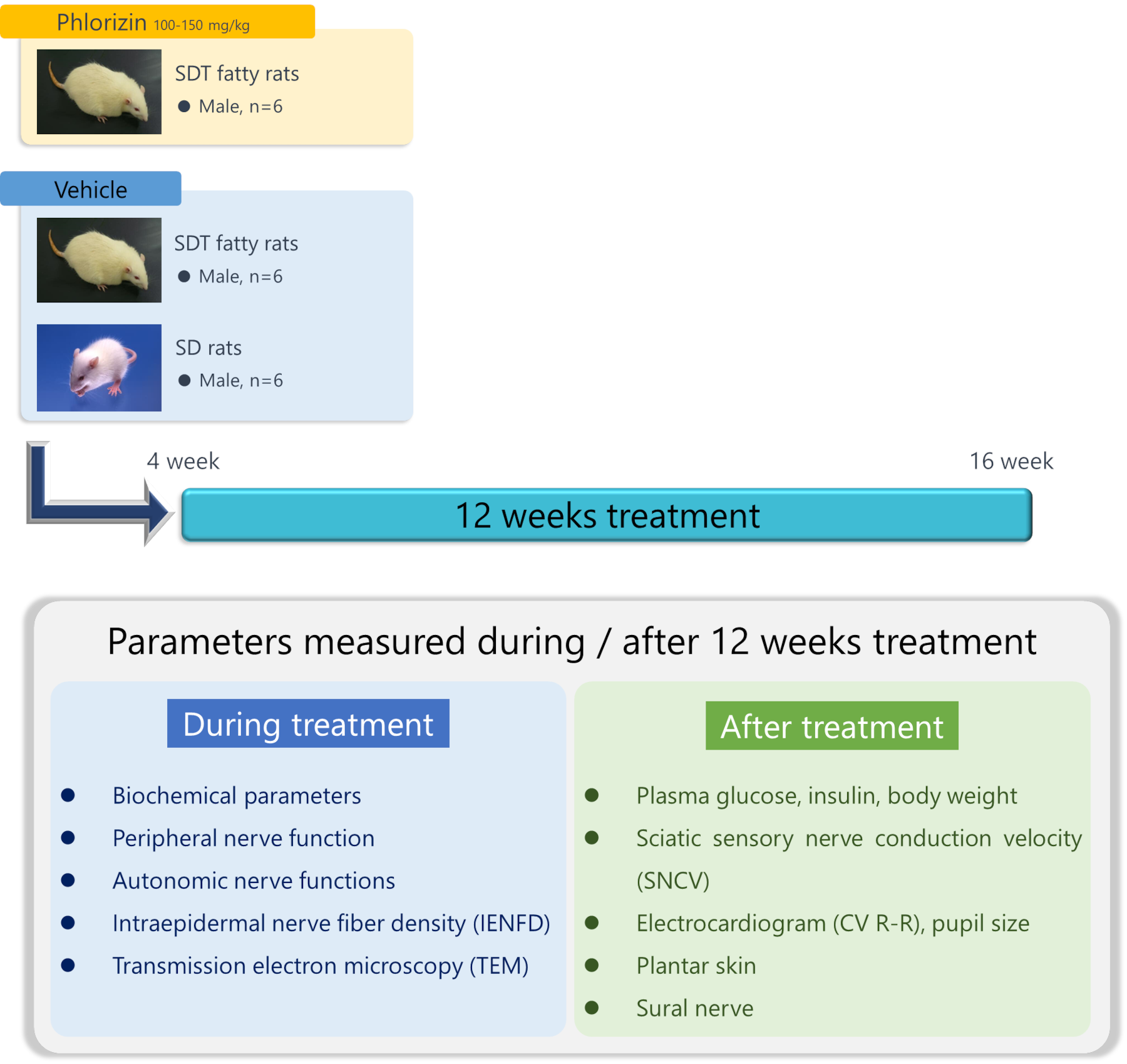
Using 4-week-old male SDT fatty rats and normal SD rats, Phlorizin 100-150mg/kg was subcutaneously administered to SDT fatty rats until 16 weeks of age.
We examined changes in blood biochemical parameters over time, measured nerve conduction velocity and evaluated autonomic nerve function at 15 weeks of age, and measured intraepidermal nerve fiber density (IENFD) and sural nerve electrons at autopsy at 16 weeks of age. Microscopic observations were performed.
Biochemical Parameters
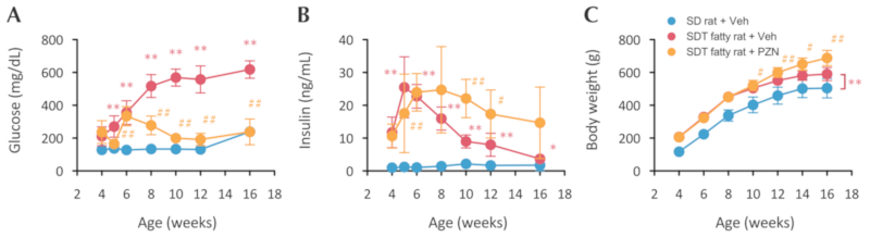
Plasma glucose levels in SDT fatty rats were significantly higher than those in age-matched SD rats. PZN treatment decreased plasma glucose levels (A). Plasma insulin levels in SDT fatty rats decreased after 5 weeks of age but were prevented by PZN treatment (B). Body weights of SDT fatty rats were significantly higher than those in SD rats through the experimental period.
PZN-treated SDT fatty rats were heavier than vehicle-treated SDT fatty rats (C). Each value represents the mean ± S.D. *p<0.05, **p<0.01 vs. SD rats. #p<0.05, ##p<0.01 vs. SDT fatty rats (Vehicle) (Two-way ANOVA with Student’s or Aspin-welch t-test).
Nerve Conduction Velocity
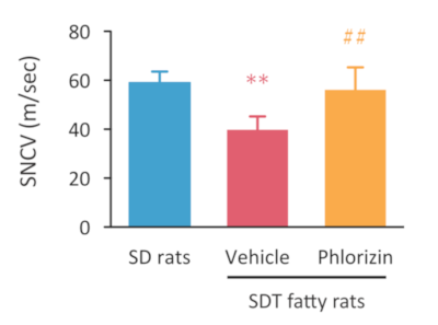
In SDT fatty rats, decreased SNCV of the sciatic nerve was improved by PZN treatment.
Each value represents the mean ± S.D. **p<0.01 vs. SD rats. ##p<0.01 vs. SDT fatty rats (Vehicle) (One-way ANOVA with Student’s or Aspin-welch t-test).
Nerve Fiber Density
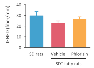
Sectioned planter skins of the hind limbs were immunostained with anti-PGP9.5 antibody. The density of nerve fiber was slightly decreased in SDT fatty rats. PZN treatment tended to prevent the decrease of IENFD. Value represents the mean ± S.D..
Autonomic Nerve Functions
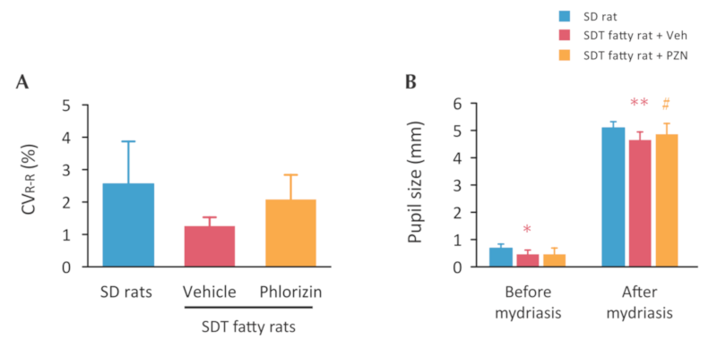
The coefficient variance of R-R (CVR-R) intervals partly decreased in SDT fatty rats. This change tended to improve by PZN treatment (A). Pupil size was smaller in SDT fatty rats both before and after instillation of mydriatic drops compared to SD rats. PZN improved the pupil size after the instillation of mydriatic drops (B). Each value represents the mean ± S.D. *p<0.05, **p<0.01 vs. SD rats. #p<0.05 vs. SDT fatty rats (Vehicle) (One-way ANOVA with Student’s or Aspin-welch t-test).
Electron Micrographs of Sural Nerve
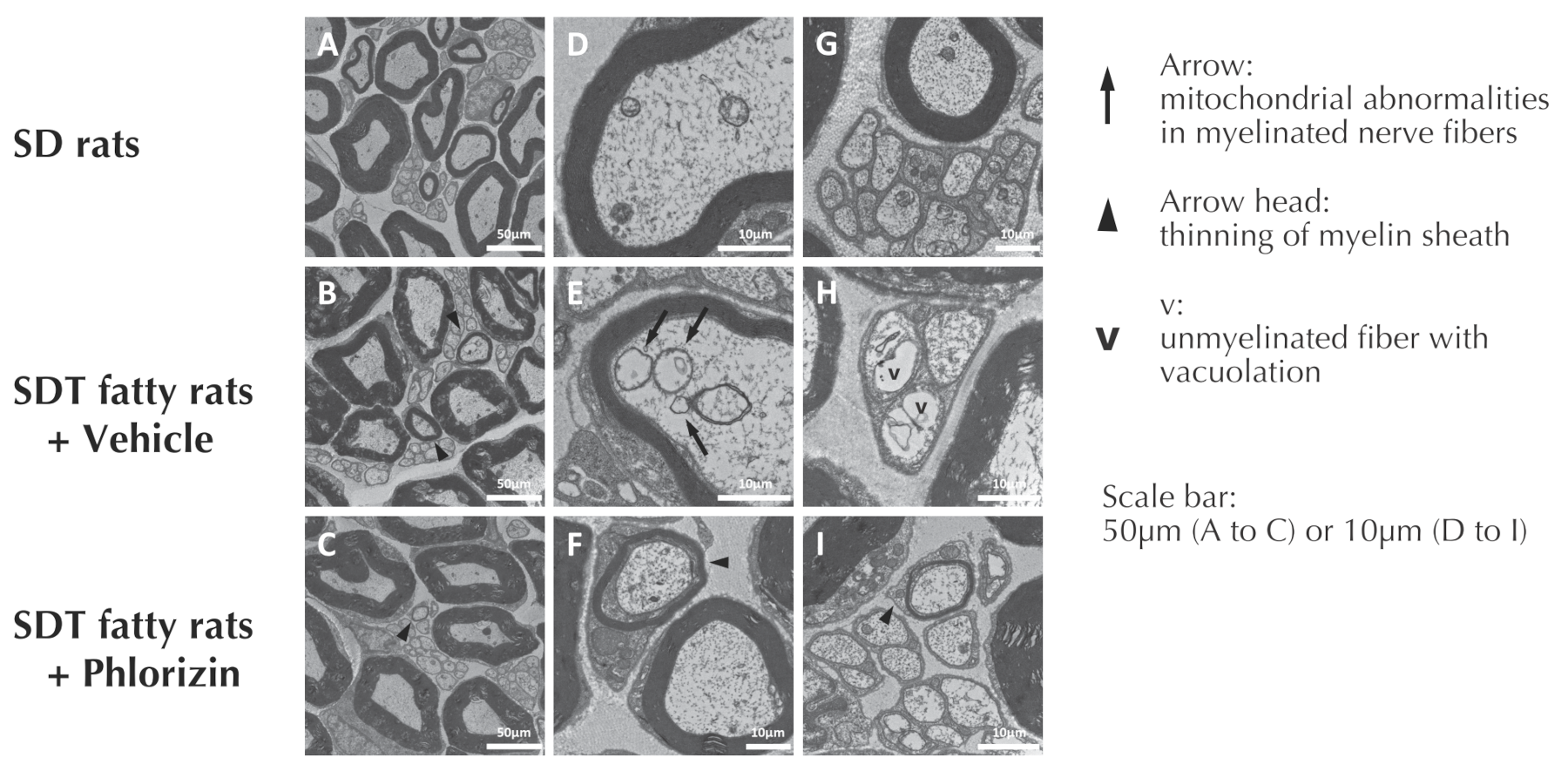
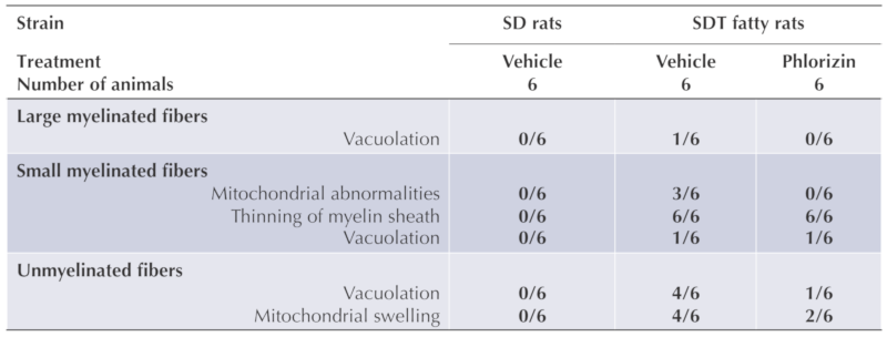
The mitochondrial abnormalities (swelling or degeneration) in small, myelinated fibers, and vacuolation and mitochondrial swelling in unmyelinated fiber were found in the SDT fatty rats. PZN treatment clearly relived those morphological abnormalities.
Conclusion
Male SDT fatty rats showed the decrease in SNCV, IENFD, and the morphological abnormalities of small myelinated and unmyelinated fibers in sural nerve by 16 weeks of age. Impaired autonomic nerve functions were also suggested. Because these abnormalities were prevented by controlling blood glucose levels, these changes seem to be caused by sustained hyperglycemia.
In addition to severe hyperglycemia, SDT fatty rats also show dyslipidemia (Matsui et al., Exp Anim, 2008) and slight hypertension (Ishii et al., Exp Anim, 2010). These multipul factors may also contribute to type 2 diabetes and its microvascular complications such as DPN. Therefore, SDT fatty rats can help future work on DPN in type 2 diabetes.
Manuscript
- Katsuda Y, Sasase T, Tadaki H, Mera Y, Motohashi Y, Kemmochi Y, Toyoda K, Kakimoto K, Kume S, Ohta T. Contribution of hyperglycemia on diabetic complications in obese type 2 diabetic SDT fatty rats: effects of SGLT inhibitor phlorizin. Experimental Animals. 2015; 64: 161-169.
- Maekawa T, Tadaki H, Sasase T, Motohashi Y, Miyajima K, Ohta T, Kume S. Pathophysiological profiles of SDT fatty rats, a potential new diabetic peripheral neuropathy model. Journal of Pharmacological and Toxicological Methods. 2017; 88: 160-166.
- Sasase T, Maekawa T, Tadaki H, Motohashi Y, Miyajima K, Ohta T, Kume S. Diabetic peripheral neuropathy in SDT fatty rat, a new animal model of obese type2 diabetes. 27th Annual Meeting of the Diabetic Neuropathy Study Group of the European Association for the Study of Diabetes (EASD) (Neurodiab 2017). Sep 9-11, 2017; Hotel Vila Galé Coimbra, Coimbra, Portugal.
Product page:
▼ SDT fatty rats product page
Inquiry
If you have any question, please feel free to contact us from below.