Diabetic retinopathy is a major complication of diabetes and a leading cause of blindness. Early detection and appropriate treatment are crucial; however, due to the complexity of the pathology, many aspects remain unclear, making the development of new treatments an urgent need.
This webpage summarizes the research findings on diabetic retinopathy using SDT fatty rats. These rats faithfully replicate the human condition and have been shown to be particularly useful for elucidating the pathology in its early stages. We present multifaceted research findings, including vascular changes, inflammatory responses, and the protective effects of pemafibrate. This research suggests new solutions for the prevention and treatment of diabetic retinopathy and is expected to contribute to addressing unmet medical needs.
Retinal capillary changes and cytokine expression in Spontaneously Diabetic Torii fatty rats
Purpose:
The prevalence of diabetes is on the rise globally. In 2021, approximately 536.6 million individuals were affected by diabetes, and this number is projected to increase to 783.2 million by 2045 [1]. One-third of adults with diabetes have diabetic retinopathy (DR), which is one of the primary causes of visual loss among those of working age [2]. Diabetic macular edema (DME) is a pathological thickening and swelling of the macula that results from microvascular complications [3]. It can develop at any stage of DR and can lead to visual impairment.
The Spontaneously Diabetic Torii (SDT) fatty rat is an animal model of obese Type 2 diabetes. The SDT fatty rats exhibit hyperglycemia within 5 weeks. [5, 6] We previously reported that the SDT fatty rats exhibit cortical and posterior subcapsular cataracts that mimic human diabetic cataracts [6].
In this study, we investigated retinopathy in the SDT fatty rats and compared their characteristics with those of human DR to clarify the similarities.
Excerpt:
Retinal thickness in SDT fatty rats and SD rats.
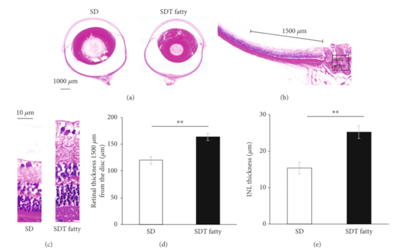
Figure 1:
Retinal thickness in SDT fatty rats and SD rats. Histopathological findings in the retina of SDT fatty rats and SD rats. (a) Representative microscopic image of hematoxylin and eosin (H&E)-stained eyeballs from 24-week-old male SD rats and SDT fatty rats. Index bar = 1000 μm. (b, c) The retinal thickness was measured at a distance of 1500 μm from the optic nerve head. (d) In SDT fatty rats, the total retinal thickness was found to be significantly greater than that in SD rats. n = 8, each. ∗∗ p < 0 01. (e) The INL thickness in SDT fatty rats was significantly higher than that in SD rats. n = 8, each. ∗∗ p < 0 01. SDT, Spontaneously Diabetic Torii; SD, Sprague–Dawley; INL, inner nuclear layer.
Pericyte/endothelial ratio in SDT fatty rats.
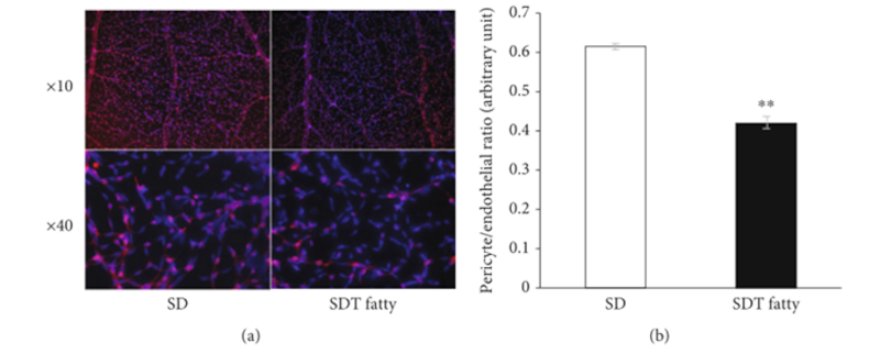
Figure 4:
Pericyte/endothelial ratio in SDT fatty rats. (a) Representative photo of the immunostained retinal vessels. NG-2 (red) and DAPI (blue). (b) The P/E ratio in SDT fatty rat retina showed a significant decrease. n = 5, each. ∗∗ p < 0 01. SDT, Spontaneously Diabetic Torii; DAPI, 4′,6-diamidino-2-phenylindole; P/E ratio, pericyte/endothelial ratio.
Conclusion:
The present study demonstrated that SDT fatty rats exhibited early diabetic changes in the retina, indicating that SDT fatty rats serve as a potential animal model in researches on the pathogenesis of early human DR.
Kasumi_K_2022_RetinalCapillaryCytokines_SDTfattyRats_Poster.pdfRetinal capillary changes and cytokine expression in Spontaneously Diabetic Torii fatty rats
Kikuchi_K_2025_SdtRat_EarlyDR_CapillaryInflammation_Paper.pdfSpontaneously Diabetic Torii Fatty Rat Shows Early Stage of Diabetic Retinopathy Characterized by Capillary Changes and Inflammation
Protective Effect of Pemafibrate Treatment against Diabetic Retinopathy in Spontaneously Diabetic Torii Fatty Rats
Purpose:
Diabetic retinopathy (DR) can cause visual impairment and blindness, and the increasing global prevalence of diabetes underscores the need for effective therapies to prevent and treat DR. Therefore, this study aimed to evaluate the protective effect of pemafibrate treatment against DR, using a Spontaneously Diabetic Torii (SDT) fatty rat model of obese type 2 diabetes.
Excerpt:
Amplitude and Peak Time of Electroretinography at 24 Weeks of Age in Each Rat Group
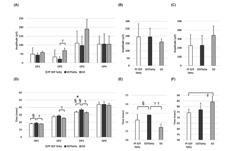
Fig.4. Amplitude and Peak Time of Electroretinography at 24 Weeks of Age in Each Rat Group
(A) Oscillatory potential (OP)1–OP4 amplitude; (B) a wave amplitude; (C) b wave amplitude; (D) OP1–OP4 peak times; (E) a wave peak time; (F) b wave peak time. No significant difference in the amplitude between the Pf SDT and SDT fatty groups was observed. In contrast, the extension of OP1 and OP3 waves during the peak time was suppressed in the Pf SDT fatty group. The data are expressed as the mean ± standard deviation. * p < 0.05, Pf SDT fatty rats vs. SDT fatty rats; # p < 0.05, Pf SDT fatty rats vs. SD rats; † p < 0.05, †† p < 0.01, SDT fatty rats vs. SD rats by Scheffe’s test. § p < 0.05, §§ p < 0.01, Pf SDT fatty rats vs. SDT fatty rats by Mann–Whitney U-test.
Retinal Thickness and Choroidal Thickness
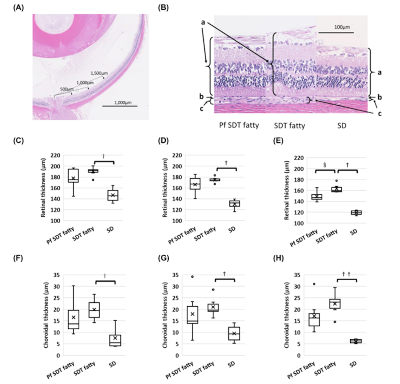
Fig. 5. Retinal Thickness and Choroidal Thickness
(A) The retinal and choroidal thicknesses were measured at 500, 1000, and 1500 microns from the optic disc. The scale bar indicates 1000 microns. (B) Comparison of the retinal and choroidal thicknesses 500 microns from the optic disc at 24 weeks of age in each rat type (hematoxylin and eosin stain). a, retina; b, retinal pigment epithelium; c, choroid. The scale bar indicates 100 microns. Retinal thicknesses at (C) 500 microns from the disc, (D) 1000 microns from the disc, and (E) 1500 microns from the disc in each rat group. The retinas at 1500 microns from the optic disc were thinner in the SDT fatty rats treated with pemafibrate (Pf SDT fatty group) than in the non-treated SDT fatty group. Choroidal thicknesses at (F) 500 microns from the disc, (G) 1000 microns from the disc, and (H) 1500 microns from the disc in each rat group. † p < 0.05, †† p < 0.01, SDT fatty rats vs. SD rats by Scheffe’s test. § p < 0.05, Pf SDT fatty rats vs. SDT fatty rats by Mann–Whitney U-test.
Conclusion:
Extension of the OP wave was suppressed in the Pf SDT fatty group in the present study. Although food consumption and blood glucose levels did not change after pemafibrate administration, the ERG data showed that pemafibrate protected retinal function. Therefore, pemafibrate is thought to be effective in suppressing the onset and progression of DR through mechanisms other than improving blood glucose levels. These results suggest that pemafibrate has a protective effect against DR.
Yishiki_T_2024_Pemafibrate_Protective_DR_SDTRat_Paper.pdfProtective Effect of Pemafibrate Treatment against Diabetic Retinopathy in Spontaneously Diabetic Torii Fatty Rats
Pathological features of diabetic retinopathy in unilaterally nephrectomized Spontaneously Diabetic Torii fatty rats given 0.3% saltwater
Purpose:
We previously reported on the Spontaneously Diabetic Torii (SDT) rat, which is an animal model of spontaneous type 2 diabetes with severe diabetic ocular complications. The SDT fatty rat, established by introducing the fa allele (obesity gene) of the Zucker fatty rat into the SDT rat genome, is a new model of obese type 2 di 1 abetes. We reported that the SDT fatty rats developed more severe diabetic retinopathy (DR) earlier than the SDT rats. It was reported recently that unilateral nephrectomy (Nx) and salt supplementation in Spontaneously Diabetic Torii (SDT) fatty rats expedite kidney disorders. The purpose is to clarify the pathological features of DR in unilaterally nephrectomized SDT fatty rats given 0.3% saltwater.
Excerpt:
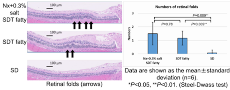
Retinal Folds
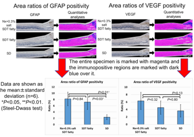
Immunostaining for GFAP and VEGF
Conclusion:
- Severe DR developed in both the Nx+0.3% salt SDT fatty and SDT fatty rats compared with the SD rats.
- However, the progression of DR in the Nx+0.3% salt SDT fatty rats was not marked compared with the SDT fatty rats.
- The progression of DR may have been suppressed by the improvement in the blood glucose level despite development of kidney dysfunction due to unilateral Nx and salt supplementation.
- Further analysis and examination are needed.
Yoshiaki_T_2019_DR_Pathology_UninephSdt_Saltwater_Poster.pdfPathological features of diabetic retinopathy in unilaterally nephrectomized Spontaneously Diabetic Torii fatty rats given 0.3% saltwater
The Uni-Nephrectomized SDT Fatty Rat, a novel Type 2 diabetic model of Diabetic Nephropathy, develops features of diabetic retinopathy over 10 weeks
Purpose:
Animal models of spontaneous diabetes are still needed to mimic human diabetic retinopathy. We recently developed a novel model of diabetic nephropathy using the uni-nephrectomized Spontaneously Diabetic Torii (SDT) fatty rat. Fed a salt rich diet, this hypertensive/obese/type 2 diabetic rat develops advanced renal glomerulosclerosis, inflammation and fibrosis, and a >50% glomerular filtration rate decline within 10 weeks. Here we investigated whether this rat model would also develop features of diabetic retinopathy.
Excerpt:
Unx SDT fatty rats show reactive gliosis in retina at 17 weeks of age
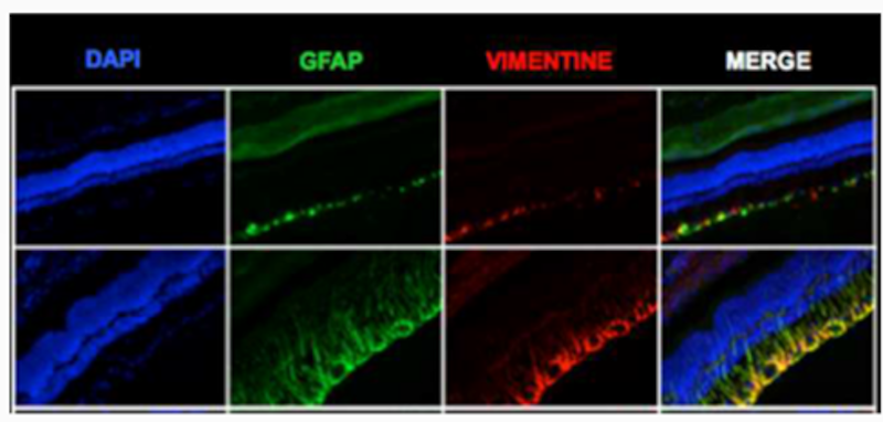
DAPI (4′,6-diamidino-2-phenylindole; for nuclear acid staining), glial fibrillary acidic protein (GFAP) and vimentin (markers of Müller cells) immunoreactivity in sagittal retinal sections, assessed by fluorescence microscopy. Upper and lower panels show representative pictures from a Unx SD rat and Unx SDT fatty rats, respectively, at 17 weeks of age.
Evans blue retinal flat-mount indicates vessel dilatation and tortuosity, without evidence of retinal vascular permeability, in 17-week old Unx SDT fatty rats
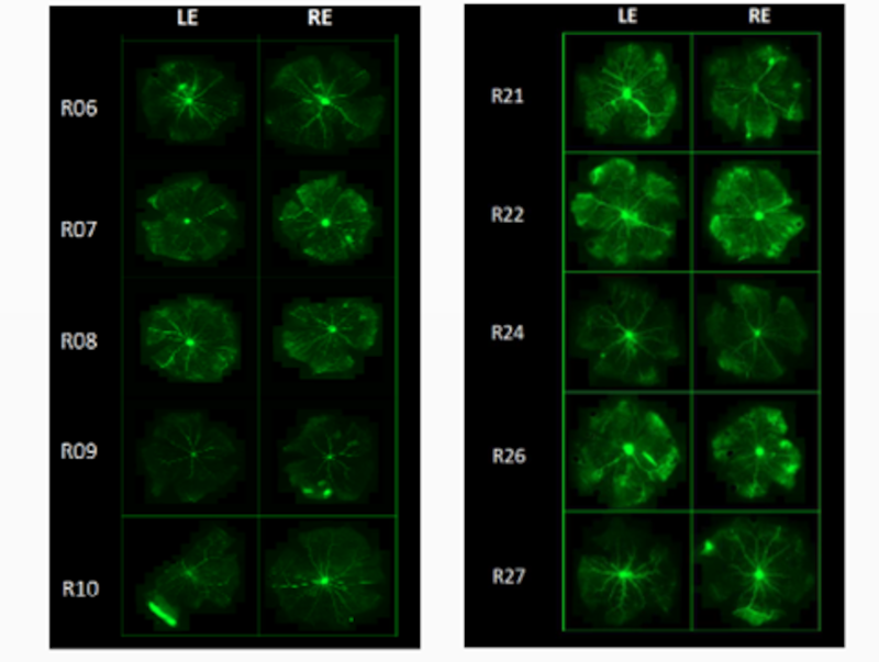
Evans blue retinal flat-mount of uni-nephrectomized SD rat (left panel) and SDT fatty rat (right panel) at 17 weeks of age.
Conclusion:
- Uni-nephrectomized SDT fatty rats develop features of diabetic retinopathy in parallel with diabetic nephropathy.
- Our original data set suggests that this rat model is suitable to evaluate the effects of drugs on these type 2 diabetes co-morbidities.
François_B_2018_UniNephSdt_DR_Model_10weeks_Poster.pdfThe Uni-Nephrectomized SDT Fatty Rat, a novel Type 2 diabetic model of Diabetic Nephropathy, develops features of diabetic retinopathy over 10 weeks
Laurence_F_2016_DR_Symptoms_Evaluation_UniNephSDT_Paper.pdfEVALUATION OF DIABETIC RETINOPATHY SYMPTOMS IN SDT FATTY RATS WITH UNILATERAL NEPHRECTOMY