Introduction
CLEA Japan offers the SDT fatty rats, a model rat that develops diabetic complications at an early stage.
Pathophysiology
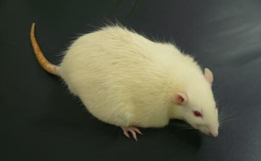
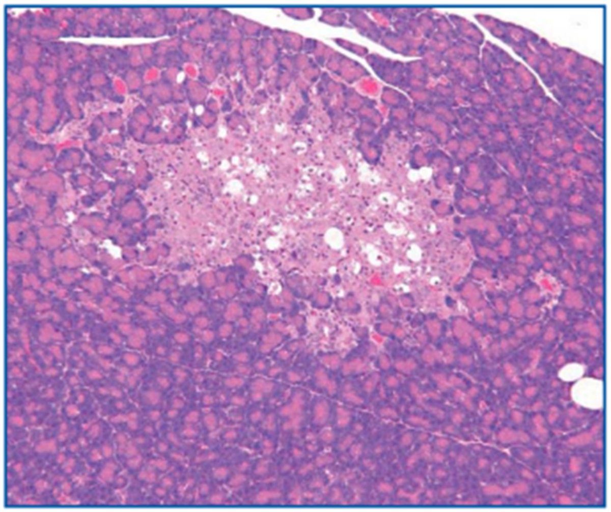
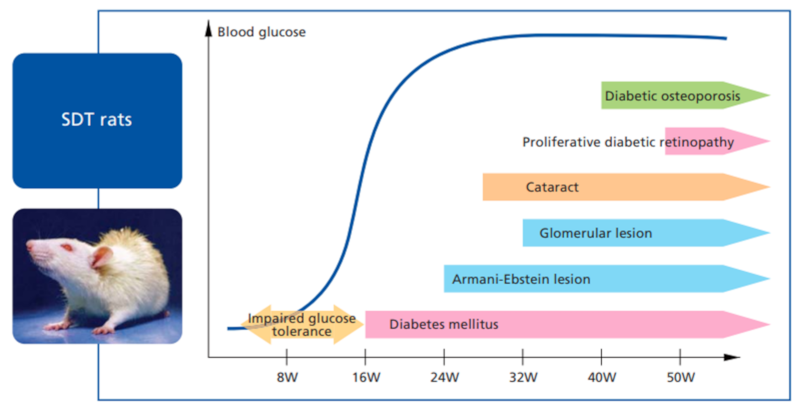
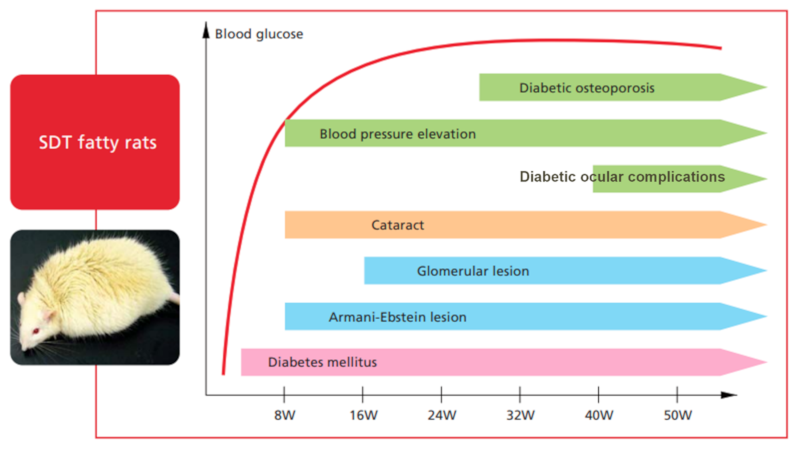
- SDT fatty rats become overtly obese just after weaning and are distinguishable from normal or original SDT rats, and show significant polyphagia. Body weights of male SDT fatty rats reach 500-600 g at 16 weeks (Figure 1). SDT fatty rats show hyperglycemia much earlier than original SDT rats and show slightly higher blood glucose level even just after weaning. Blood glucose level reaches 600 mg/dl at 8 weeks of age, and 800 mg/dl at 12 weeks and thereafter (Figure 2). Hyperlipidemia is also observed in SDT fatty rats2). Blood insulin levels increase at 4-8 weeks of age and decrease thereafter (Figure 3). Histopathologically, hypertrophy, vacuolation, and irregular shape in pancreatic islet are found (Figure 4, 13 weeks).
- In addition, computerized tomography (CT) shows excess accumulation of abdominal /subcutaneous fat. Noticeable fatty liver is also observed in SDT fatty rats4). These features make SDT fatty rat a highly obese model associated with disorder of lipid metabolism. Furthermore, increased diastolic blood pressure6) (from 8 weeks of age) suggests use of SDT fatty rats as model of metabolic syndrome. All of these pathophysiological observations are found in both male and female SDT fatty rats5)
Figure-1 . Changes in body weight of male SDT fatty rats (*P<0.05, ** P<0.01)
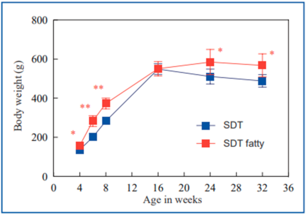
Figure-2 . Changes in nonfasting plasma glucose of male SDT fatty rats (*P<0.05)
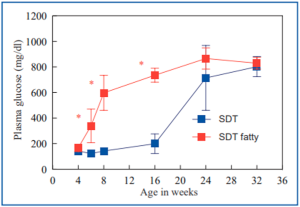
Figure-3 . Changes in nonfasting plasma insulin of male SDT fatty rats (*P<0.05, ** P<0.01)
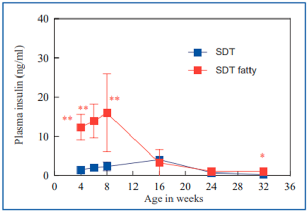
Figure-4 . Histopathological findings in pancreas of male SDT fatty rat (13 weeks of age)
Notes on rearing and handling SDT fatty rats
- Diet and drinking water
- High-volume bedding and frequent cage-change are recommended
Background and Origin
In 2004, Dr. Masuyama and Dr. Shinohara (Research Laboratories of Torii Pharmaceutical Co., Ltd., Japan) established the congenic type 2 diabetes model Spontaneously Diabetic Torii fatty (SDT fatty) rat by introducing the fa allele of the Zucker Fatty rat into the genome of the original SDT rat1). SDT fatty rats were introduced to Central Pharmaceutical Research Institute, Japan Tobacco Inc. (Japan) and were characterized in detail by Dr. Ohta and Dr. Sasase. CLEA Japan has received right of production and sales from Japan Tobacco Inc., and has distributed the animals as SDT fatty rats from 2012.
Diabetic ocular complications
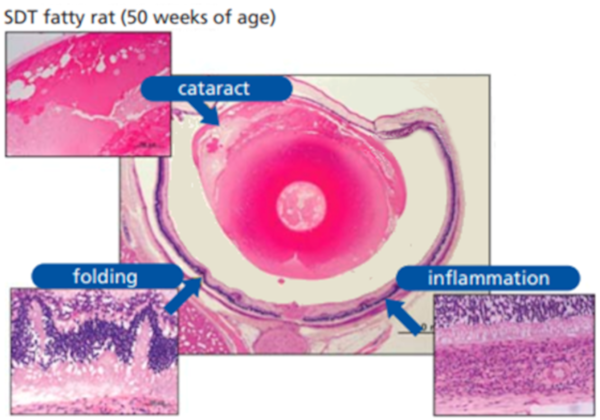
With elevated glucose level from young age, macroscopic opacity occurs in the SDT fatty rat from 8 weeks of age and progresses severely to mature cataract in almost all animals by 16 weeks of age2). Retinal lesions (retinal folding and thickening) and inflammation in the choroid (uveitis) are observed in SDT fatty rats at 50 weeks of age (Figure 5) 17). Uveitis associated with diabetes has been reported in humans; however, there has been no report of a spontaneous diabetes animal model affected with uveitis. SDT fatty rats after 16 weeks of age show a prolongation of the peak latency on an electroretinogram2). On the other hand, fibrous proliferation and following tractional retinal detachment observed in the original SDT rats are not observed in SDT fatty rats up to 60 weeks of age.
Figure-5 . Histopathological findings in ocular of female SDT fatty rat (50 weeks of age)
Diabetic nephropathy
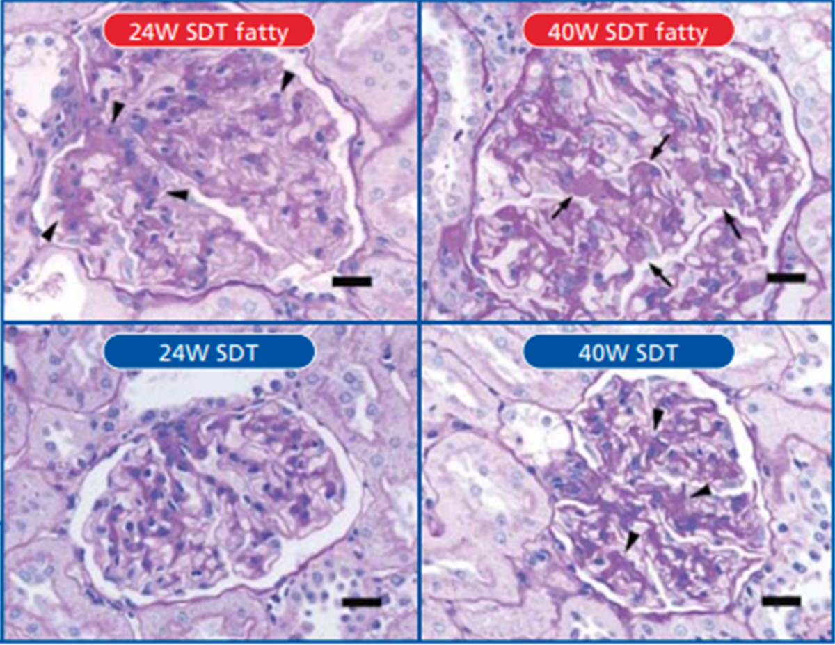
SDT fatty rats show marked polyuria accompanied by glucosuria and proteinuria due to persistent hyperglycemia from early age. Histopathologically, glycogen deposition in the renal tubule (Armanni-Ebstein change) and tubular dilatation caused by polyuria are observed from 8 weeks of age. In the glomerulus, increased mesangial matrix and diffuse glomerulosclerosis are observed, and the nodular-like lesion appears in the mesangium from approximately 40 weeks of age (Figure 6). Tubulointerstitial lesions including fibrosis, inflammatory cell infiltration, and regeneration of tubules are observed after 50 weeks of age18).
Figure-6 . Histopathological findings in kidney of male SDT fatty rat (24,40 weeks of age)
Diabetic neuropathy
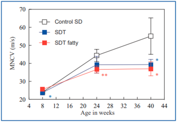
SDT fatty rats show diabetic peripheral neuropathy concomitant with severe hyperglycemia. In an electrophysiological study, the caudal motor nerve conduction velocity (MNCV) in SDT rats was decreased at 24 weeks of age and it became clear with age (Figure 7). Decreased caudal blood flow and excess sorbitol accumulation in sciatic nerve were also observed. Morphologically, the number of sural nerve fibers was lower at 40 weeks of age, and occluded/thickened epineurial arterioles were frequently found in SDT fatty rats19). SDT fatty rats are susceptible to diabetic diarrhea, suggesting development of diabetic autonomic neuropathy.
Figure-7 . Changes in MNCV of male SDT fatty rats (*P<0.05, ** P<0.01)
Diabetic osteoporosis
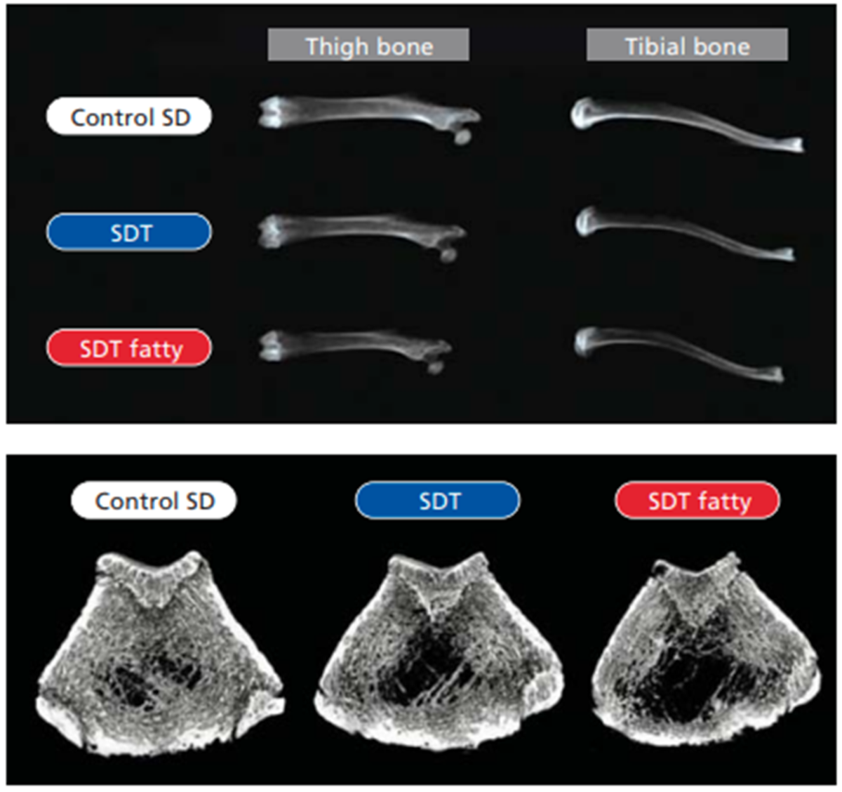
SDT fatty rats show osteoporosis with low bone turnover, unlike an ovariectomized model. Levels of serum osteocalcin, a bone formation marker, and urine deoxypyridinoline (DPD), a bone resorption marker, are decreased from 8 weeks of age in SDT fatty rats (Figure 8). At 40 weeks of age, SDT fatty rats show low bone mineral density (BMD), shortening of bone length, and deterioration in trabecular bone microstructure (Figure 9) of the long bones. Furthermore, high serum homocysteine levels in SDT fatty rats at 32 weeks of age suggest deterioration in bone quality. Parathyroid hormone (PTH) injections suppress a decrease in BMD and also decrease serum glucose levels in SDT fatty rats.
Figure-8 . Metabolism parameter of male SDT fatty rats (*P<0.05, ** P<0.01)
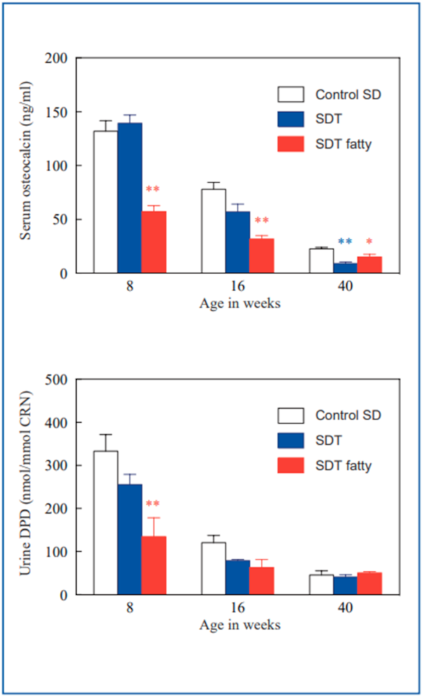
Figure-9 . Computed tomography images male SDT fatty rats (40 weeks of age)
Pharmacological treatment
Several anti-diabetic drugs such as biguanide (metformin), thiazolidinedione (pioglitazone), and DPPIV inhibitors improve hyperglycemia in SDT fatty rats. Controlling blood glucose level from early stage can retard progression of insulin hyposecretion9) and diabetic complications, such as diabetic peripheral neuropathy19).
Conclusion
In addition to early development of hyperglycemia, multiple features such as obesity, hypertension, and osteoporosis make SDT fatty rat a useful model for diabetes, diabetic complications, and metabolic syndromes. The characteristics of SDT fatty rat are also suitable for use in drug discovery of these diseases
Comparison of the pathology of SDT rats DT rats and SDT fatty rats
References
1) Masuyama T, Katsuda Y, and Shinohara M: A novel model of obesity-related diabetes: introgression of the Leprf@ allele of the Zucker fatty ratin to nonobese Spontaneously Diabetic Torii (SDT) rats. Exp. Anim. 54(1):13-20, 2005
https://pubmed.ncbi.nlm.nih.gov/15725677/
2) Matsui K, Ohta T, Oda T, Sasase T, Ueda N, Miyajima K, Masuyama T, and Matsushita M: Diabetes-associated complications in Spontaneously Diabetic Torii fatty Rats. Exp. Anim. 57 (2): 111-121, 2008 (2008 Japanese Society of Animal Technology Award for Best Paper)
https://pubmed.ncbi.nlm.nih.gov/18421173/
3) Matsui K, Ohta T, Morinaga H, Sasase T, Fukuda S, Ito M, Ueda M, Ogawa N, Miyajima K, and Matsushita M: Effects of preventing hyperphagia on glycolipid metabolic abnormalities in Spontaneously Diabetic Torii fatty Rats. Anim. Sci. J. 79 (5) 605-613, 2008 (Paper awarded the 2010 Animal Science Journal Excellent Paper Award)
Effects of preventing hyperphagia on glycolipid metabolic abnormalities in Spontaneously Diabetic Torii fatty Rats - Google 検索
4) Morinaga H, Ohta T, Matsui K, Sasase T, Fukuda S, Ito M, Ueda M, Ishi Y, Miyajima K, and Matsushita M: Effects of food restriction on adipose tissue in Spontaneously Diabetic Torli fatty Rats. Exp. Diabetes Res. 2009:715057. 2009.
https://pubmed.ncbi.nlm.nih.gov/19696902/
5) Ishii Y, Ohta T, Sasase T, Morinaga H, Ueda N, Hata T, Kakutani M, Miyajima K, Katsuda Y, Masuyama T, Shinohara M, and Matsushita M: Pathophysiological analysis of female Spontaneously Diabetic Torii fatty Rats. Exp. Anim. 59 (1): 73-84, 2010.
https://pubmed.ncbi.nlm.nih.gov/20224171/
6) Ishii Y, Maki M, Yamamoto H, Sasase T, Kakutani M and Ohta T: Evaluation of blood pressure in Spontaneously Diabetic Torii-Lepr(fa) rats. Exp. Anim. 59 (4): 525-529, 2010.
https://pubmed.ncbi.nlm.nih.gov/20661000/
7) Ishii Y, Ohta T, Sasase T, Morinaga H, Miyajima K, and Kakutani M: Effect of food restriction on pancreatic islet in Spontaneously Diabetic Torii fatty rats. J. Vet. Med. Sci. 73(2): 169-175, 2011.
https://pubmed.ncbi.nlm.nih.gov/20877155/
8) Ishii Y, Maki M, Yamamoto H, Sasase T, Kakutani M, and Ohta T: Blood pressure characteristics of female Spontaneously Diabetic Torii Lepr rats. J. Vet. Med. Sci. 73(4): 501-505. 2011.
https://pubmed.ncbi.nlm.nih.gov/21127391/
9) Fukuda S, Miyajima K, Sasase T and Ohta T: Spontaneously Diabetic Torii Lepr? (SDT fatty) Rats — A novel model of obese type2 diabetes -.Open Diabetes J. 4: 30-36, 2011.
Spontaneously Diabetic Torii Lepr? (SDT fatty) Rats — A novel model of obese type 2 diabetes - Google 検索
10) Kimura S, Sasase T, Ohta T, Sato E, and Matsushita M: Characteristics of bone turnover, bone mass and bone strength in Spontaneously Diabetic Torii- Lepr? rats. J. Bone Miner. Metab. (in press).
https://pubmed.ncbi.nlm.nih.gov/22038286/
11) Kimura S, Sasase T, Ohta T, and Matsushita M: Effects of ovariectomy on bone metabolism and bone mineral density in Spontaneously Diabetic Torii Lepr(fa) rats. J. Vet. Med. Sci. 73(8): 1025-1029, 2011.
https://pubmed.ncbi.nlm.nih.gov/21490414/
12) Tokumone N, Inaba N, Nakamura S, Hirao N, Maeno T, Chiba K, Sasase T, and Ota T: Cryopreservation of 2-cell stage embryos of the spontaneously diabetic Torii-Lepr(fa) (SDT-fa/fa) rat using a simplified vitrification method 46(1):3-9,2011. 31st Research Encouragement Award from the Japanese Association of Laboratory Animal Technicians
Cryopreservation of 2-cell stage embryos of the spontaneously diabetic Torii-Leprfa (SDT-fa/fa) rat using a simplified vitrification method - Google 検索
13) Kimura S, Sasase T, Ohta T, Sato E, and Matsushita M: Parathyroid hormone (1-34) improves bone mineral density and glucose metabolism in Spontaneously Diabetic Torii- Lepr(fa) rats. J. Vet. Med. Sci. 74(1): 103-105, 2012.
https://pubmed.ncbi.nlm.nih.gov/21869570/
14) Inaba N, Nakamura S, Hirao N, Tokumone N, Chiba K, Mitani K, and Ota T: Reproductive characteristics of female spontaneously diabetic Torii-Leprfa (SDT-fa/fa) rats
Reproductive characteristics of female spontaneously diabetic Torii-Leprfa (SDT-fa/fa) rats - Google 検索
15) Hata T, Ohta T, Ishii Y, Sasase T, Yamaguchi T, Mera Y, Miyajima K, Tanoue G, Sato E, and Matsushita M : Elevated glucagon-like peptide-1 on a high-fat diet feeding prevents the incidence of diabetes mellitus in Spontaneously Diabetic Torii- Leprf rats. J. Diabetes Mellitus (in press).
Elevated glucagon-like peptide-1 on a high—fat diet feeding prevents the incidence of diabetes mellitus in Spontaneously Diabetic Torii- Leprf rats. J. Diabetes Mellitus (in press). - Google 検索
16) Hirao N, Tokumone N, Inaba N, Chiba K, Sasase T, and Ota T: Effects of two types of basal diets (CRF-1 and CE-2) on the development and progression of diabetes in SDT-fa/fa and SDT-+/+ rats Experimental Animals
Effects of two types of basal diets (CRF-1 and CE-2) on the development and progression of diabetes in SDT-fa/fa and SDT-+/+ rats Experimental Animals - Google 検索
17) Kenmochi Y, Mitani K, Yasui Y, Masuyama T, Ota T, Sasase T, and Kakimoto T: Spontaneous uveitis in spontaneously diabetic Torii-Leprfa (SDT-fa/fa) rats: A histopathological study The 26th Annual Meeting of the Japanese Society of Diabetic Complications Omiya 2011.
Spontaneous uveitis in spontaneously diabetic Torii-Leprfa (SDT-fa/fa) rats - Google 検索
18) Kenmochi Y, Miyajima K, Yasui Y, Masuyama T, Shoda T, Toyoda K, Takahashi A, Ohta T, Sasase T, and Kakimoto K: Severely Progressed Diabetes-Assoclated Renal Complication in the Spontaneously Diabetic Torii (SDT) Fatty Rat at Ages over 50 Weeks - Histopathological Examination -. Society of Toxicologic Pathology, 31% Boston, 2012.
Severely Progressed Diabetes-Assoclated Renal Complication in the Spontaneously Diabetic Torii (SDT) Fatty Rat at Ages over 50 Weeks - Google 検索
19) Yamaguchi T, Sasase T, Mera Y, Tomimoto D, Ohta T, Sato E, and Matsushita M: Diabetic peripheral neuropathy in Spontaneously Diabetic Torii – Lepr(fa) (SDT fatty) rats. J. Vet. Med. Sci. (in press).
https://pubmed.ncbi.nlm.nih.gov/22850562/
Inquiry
If you have any question, please feel free to contact us from below.