Introduction
CLEA Japan offers the SDT/Jcl Rats, a type 2 diabetes model that develops Diabetic Ocular Complications.
Background and Origin 1)
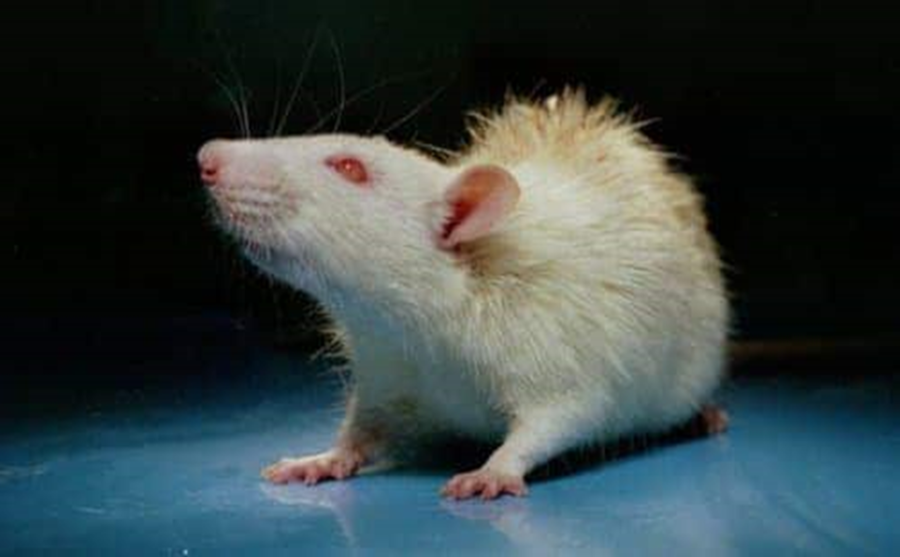
- In 1988, twelve-month-old male rats that exhibited polydipsia, polyphagia, polyuria, and glucosuria were found in Sprague-Dawley rats by Shinohara at the Research Laboratories of Torii Pharmaceutical Co., Ltd., Japan. They were mated with normal female rats of the same strain to maintain the disease symptoms and in 1991 several rats were found to exhibit positive urinary glucose earlier (at four to five months of age). An inbred strain of a non-obese type 2 diabetic model was established by repeating this sisterbrother mating and the animals obtained were named the Spontaneously Diabetic Torii (SDT) rats in 1997. The SDT rats have diabetic ocular complications such as cataracts, proliferative retinopathy, and neovascularized glaucoma, after exhibiting the primary symptoms of diabetes. Therefore, they are expected to be especially useful for research on diabetic complications and drug development for diabetes. CLEA Japan obtained the strain at F24 from Torii Pharmaceutical Research Laboratories in 1998 and started to distribute the animals as SDT/Jcl rats from 2005 (F47 as of April 2005). *Cover photograph: Appearance of a diabetic male SDT rat ages 35 weeks.
Notes on Breeding and Experimentation
- Onset of Diabetes and Clinical Characteristics of SDT rats1)
- Body Weight and Body Mass Index (BMI)1, 2, 4)
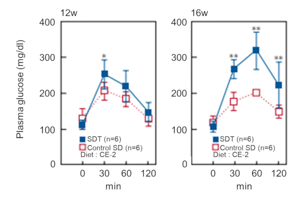
- Oral Glucose Tolerance Test (OGTT) 1, 2, 4, 5)
Figure-1 . Cumulative incidence of diabetes in SDT rats
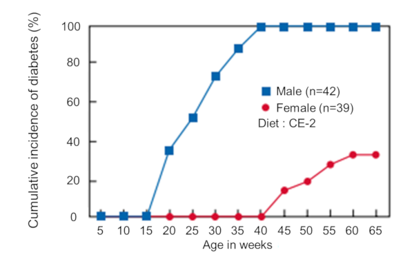
Figure-2 . Plasma glucose levels of male SDT rats in Non-fasting period (*P<0.05, ** P<0.01)
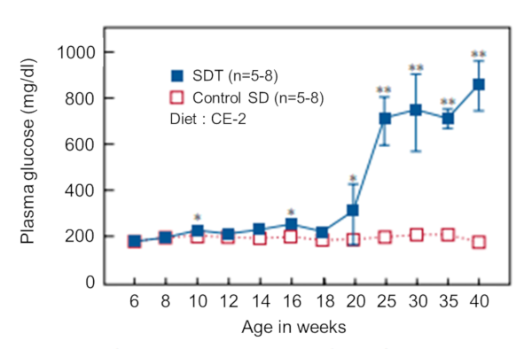
Figure-3 . Body weight curves of male SDT rats (*P<0.05, ** P<0.01)
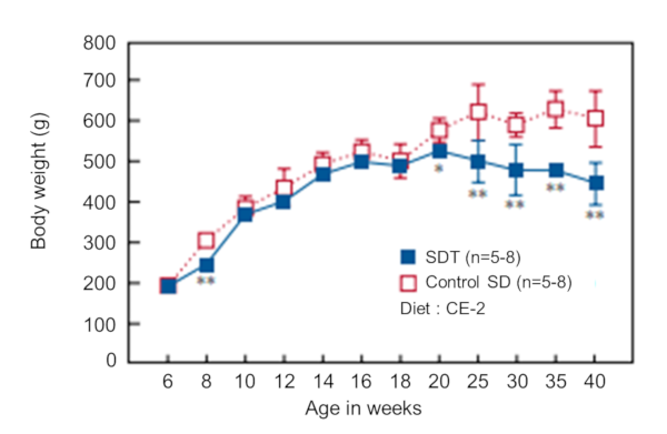
Figure-4 . Plasma glucose levels of male SDT rats (12 weeks of age, 16 weeks of age) from OGTT (*P<0.05, ** P<0.01)
- Genetic Analysis 3)
- Histopathological Examinations of the Pancreas 1, 2, 4, 5)
Figure-5 . Histopathological findings of the pancreas of a male SDT rats (25 weeks of age)
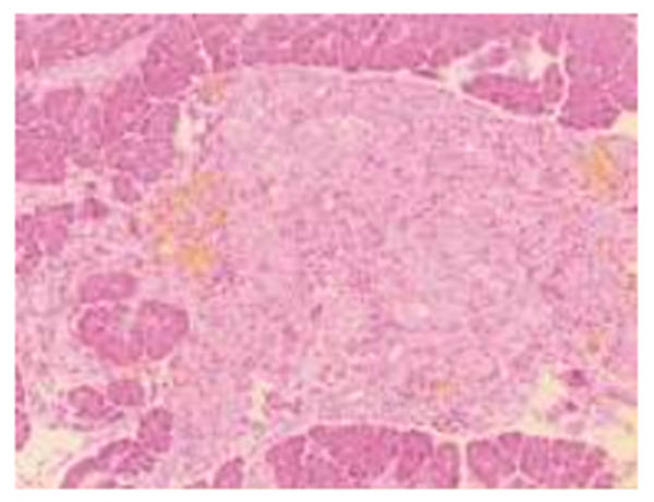
Figure-6 . Histopathological findings of the lens of a male SDT rat (40 weeks of age)
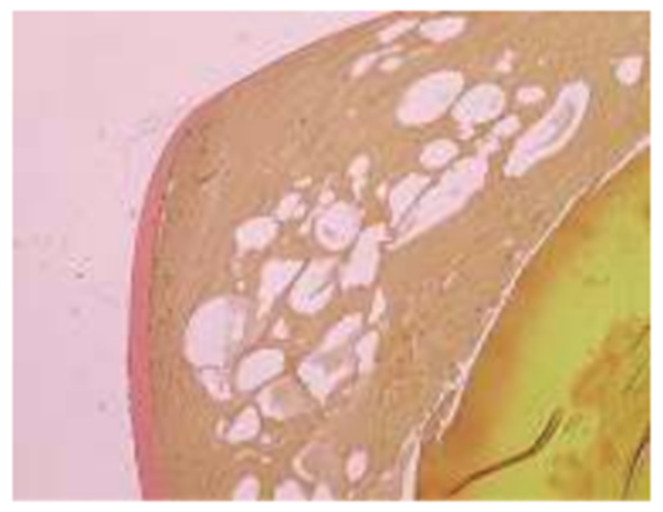
Figure-7 . Fluorescein angiography findings of the ocular of a male SDT rat (58 weeks of age)
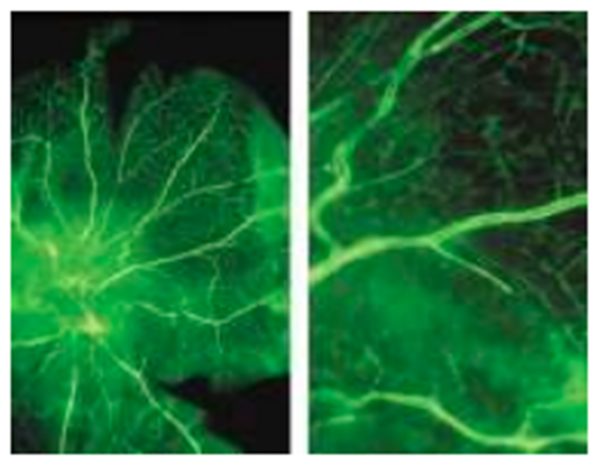
Figure-8 . Histopathological findings of the retina of a male SDT rat (70 weeks of age)
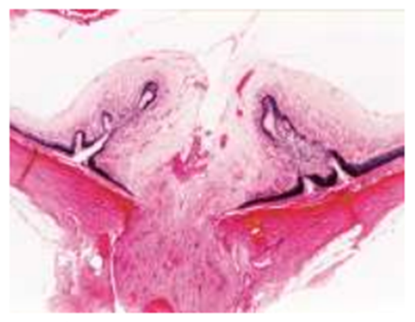
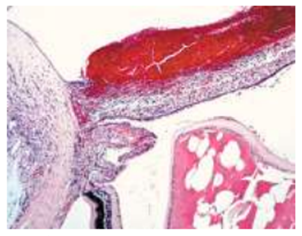
- Diabetic Ocular Complications 1, 6, 7, 8, 9)
Figure-9 . Histopathological findings of the iris and Surrounding area of a male SDT rat (77 weeks of age)
References
1) Shinohara M, Masuyama T, Shoda T, Takahashi T, Katsuda Y, Komeda K, Kuroki M, Kakehashi A, Kanazawa Y. A new spontaneously diabetic non-obese Torii rat strain with severe ocular complications. Int J Exp Diabetes Res. 2000; 1: 89- 100.
https://pubmed.ncbi.nlm.nih.gov/11469401/
2) Masuyama T, Komeda K, Hara A, Noda M, Shinohara M, Oikawa T, Kanazawa Y, Taniguchi K. Chronological characterization of diabetes development in male Spontaneously Diabetic Torii rats. Biochem Biophys Res Commun. 2004; 314: 870-7.
https://pubmed.ncbi.nlm.nih.gov/14741717/
3) Masuyama T, Fuse M, Yokoi N, Shinohara M, Tsujii H, Kanazawa M, Kanazawa Y, Komeda K, Taniguchi K. Genetic analysis for diabetes in a new rat model of nonobese type 2 diabetes, Spontaneously Diabetic Torii rat. Biochem Biophys Res Commun. 2003; 304: 196-206.
https://pubmed.ncbi.nlm.nih.gov/12705906/
4) Shinohara M, Shoda T, Oikawa T, Masuyama T, Takahashi T, Katsuda Y, Komeda K, Sato K, Kanazawa Y. Diabetic features of female SDT rats with spontaneous type 2 diabetes mellitus. J. Japan Diab. Soc. 2004; 47: 111-6. (in Japanese)
Diabetic features of female SDT rats with spontaneous type 2 diabetes mellitus - Google 検索
5) Shinohara M, Oikawa T, Sato K, Kanazawa Y. Glucose intolerance and hyperlipidemia prior to diabetes onset in female Spontaneously Diabetic Torii (SDT) rats. Experimental Diab Res. 2004; 5: 253-6.
https://pubmed.ncbi.nlm.nih.gov/15763939/
6) Kakehashi A, Kanazawa Y. SDT rat: A new diabetic retinopathy animal model. Endocrinology & Diabetology. 2001; 12: 386-90. (in Japanese)
A new diabetic retinopathy animal model. Endocrinology & Diabetology - Google 検索
7) Kakehashi A. Pathogenesis of diabetic retinopathy: Lessons from diabetic animal model. Endocrinology & Diabetology. 2003; 17: 31-7. (in Japanese)
Pathogenesis of diabetic retinopathy: Lessons from diabetic animal model. Endocrinology & Diabetology. 2003; 17: 31-7. (in Japanese) - Google 検索
8) Kakehashi A, Saito Y, Mori K, Sugi N, Ono R, Yamagami H, Shinohara M, Tamemoto H, Ishikawa S, Kawakami M, Kanazawa Y. Characteristics of diabetic retinopathy in SDT rats. Diabetes Metab Res Rev. 2005; in press.
https://pubmed.ncbi.nlm.nih.gov/16572493/
9) Yamada H, Yamada E, Higuchi A, Matsumura M. Retinal neovascularisation without ischemia in the Spontaneously Diabetic Torii rat. Diabetologia. 2005; in press.
https://pubmed.ncbi.nlm.nih.gov/15977012/
Notes on rearing and handling SDT rats
- Diet and drinking water In SDT rats, glucosuria appears on ingestion of standard diet for mice and rats. Their diet intake increases (about 1.5 to 2 times that of SD strain rats) and water intake also significantly increases (200 to 400 ml/day and more) with the onset of diabetes. It is, therefore, important to always have sufficient amounts of diet and drinking water.
- Frequent change of bedding and cleaning of cages are recommended The SDT rats urinate a lot and their bedding easily becomes dirty after onset of diabetes. Therefore, they can easily develop urinary tract infections. In order to avoid such infections, each rat should be reared alone and a large amount of bedding, with frequent replacement, should be used with plastic cage rearing. It is also recommended to change bedding frequently with bracket cage rearing.
Inquiry
If you have any question, please feel free to contact us from below.